Immediate grayish-white blanching is a warning sign, not the desired endpoint, during vascular laser treatment. It indicates that laser-generated heat has likely spread beyond the target blood vessel and caused thermal coagulation of proteins in surrounding dermal or epidermal tissue. Because the whitening is nonspecific, it does not demonstrate selective vessel destruction; it suggests that adjacent tissue is also being injured.
In vascular laser procedures, immediate gray-white blanching usually means excessive or poorly confined thermal injury. The treatment should be reassessed immediately rather than continued toward a whiter appearance, because the same heat diffusion can progress to blistering, necrosis, pigmentary change, and scarring.
Why the Skin Turns Grayish-White
Heat denatures tissue proteins
Vascular lasers are absorbed primarily by hemoglobin within the target vessel. If the delivered heat is excessive, or remains in tissue too long, it conducts outward into the vessel wall and surrounding connective tissue.
That heat can denature and coagulate dermal proteins. The resulting change in tissue structure alters light scattering, producing an immediate gray, white, or porcelain-like appearance.
The reaction is not vessel-specific
Selective photothermolysis aims to confine thermal injury to the vessel by matching the wavelength, fluence, pulse duration, and cooling strategy to the target.
Blanching indicates that this confinement may have failed. The visible response therefore reflects broad tissue coagulation rather than a specific indication that the abnormal vessel alone has been adequately treated.
Epidermal injury may also contribute
When fluence is too high or epidermal cooling is inadequate, energy can overheat the epidermis. This is particularly important in darker skin types, where epidermal melanin can compete with hemoglobin for laser energy and increase superficial heating.
Gray-white change may therefore signal combined injury to perivascular dermis and overlying epidermis, not merely successful vascular photothermolysis.
Why Blanching Became an Unreliable Endpoint
Early treatments often depended on visible whitening
Older continuous-wave and long-pulse vascular laser approaches sometimes used blanching as a clinical endpoint. The assumption was that a progressively white vessel or treatment area represented sufficient thermal destruction.
In practice, the endpoint could require excessive energy delivery and produce collateral damage before the vessel was selectively treated.
The clinical consequences can be delayed
Immediate whitening may be followed by pain, blistering, epidermal breakdown, prolonged weeping, and later textural or pigmentary changes.
Historical treatment approaches that pursued this endpoint were associated with substantial hypertrophic-scarring rates, reported in the primary reference as approximately 9% to 26%.
Selective treatment uses controlled tissue effects
Modern protocols generally aim for vessel-specific thermal injury rather than indiscriminate whitening. Pulse duration is selected in relation to vessel size so that the vessel receives sufficient thermal exposure while surrounding tissue has a better opportunity to dissipate heat.
The desired response depends on the device, wavelength, vessel, skin type, and treatment setting. Gray-white blanching should not be treated as a universal success criterion.
How to Distinguish It From Other Laser Responses
Blanching is different from transient vasoconstriction
A temporary reduction in redness can occur because of vasoconstriction or immediate vascular response. That is not necessarily the same as a fixed gray-white change in the treated skin.
The concerning pattern is immediate, tissue-level whitening or graying that suggests coagulation and excessive heating.
It is not primarily a photoacoustic pigment endpoint
A photoacoustic response associated with pigment-targeting procedures should not be used to explain vascular-laser blanching. In vascular treatment, the relevant mechanism is predominantly thermal energy absorption and heat diffusion, not a pigment micro-explosion.
Confusing these mechanisms can lead to inappropriate interpretation of the treatment endpoint.
Understanding the Trade-offs
More energy does not necessarily improve vessel clearance
Increasing fluence may increase thermal injury without producing proportionally better vascular clearance. Once heat spreads beyond the vessel, the risk-benefit balance shifts toward epidermal and dermal damage.
The objective is not to maximize visible whitening; it is to achieve adequate target-vessel injury with the least unnecessary collateral heating.
Wavelength selection matters
Some wavelengths are absorbed by both oxyhemoglobin and epidermal melanin. In heavily pigmented skin, this can reduce energy reaching deeper vessels while increasing epidermal heating.
Appropriate wavelength selection, conservative parameter adjustment, and effective epidermal cooling are therefore essential for reducing blistering and pigmentary complications.
Cooling is protective but not a substitute for correct parameters
Active cooling can reduce epidermal temperature and improve the safety margin. It cannot fully compensate for an excessive fluence, an unsuitable pulse duration, poor coupling, or an incorrectly selected wavelength.
Cooling should support—not replace—careful treatment planning.
What to Do When Immediate Blanching Appears
Stop and reassess
If grayish-white blanching appears unexpectedly or extends beyond the intended vessel, treatment should be stopped at that site. Continuing to deliver pulses risks converting reversible thermal stress into deeper tissue injury.
The area should be evaluated for pain, texture change, blistering, and signs of epidermal compromise.
Cool the treated area promptly
Appropriate cooling, such as an ice-based method suitable for the clinical setting or the device’s active cooling system, can help limit ongoing thermal injury.
Any subsequent wound care should follow the practitioner’s established protocol, particularly if blistering or epidermal disruption develops.
Use test sites and early-pulse observation
Test spots on untreated skin help reveal how the patient’s skin and target vessel respond before treating a larger area. Early pulses should be observed closely rather than assuming that a standard setting will be safe for every patient.
Making the Right Choice for Your Goal
The practical interpretation should be based on the treatment objective, skin type, vessel characteristics, and the specific laser platform.
- If your primary focus is vessel clearance: Use parameters designed for selective photothermolysis and do not pursue immediate gray-white blanching as the endpoint.
- If your primary focus is epidermal safety: Stop when unexpected blanching occurs, cool the area promptly, and reassess fluence, pulse duration, wavelength, and cooling.
- If your primary focus is treating darker skin types: Account for competing epidermal melanin absorption and use conservative, individually tested settings.
- If your primary focus is preventing scarring: Avoid cumulative thermal injury, monitor the first pulses carefully, and treat blanching as a warning of nonspecific damage.
The safest vascular laser endpoint is controlled target-vessel treatment—not maximal visible whitening.
Summary Table:
| Sign | Meaning | Risk |
|---|---|---|
| Immediate grayish-white blanching | Non-specific thermal coagulation of dermal and epidermal tissue | Blistering, necrosis, pigmentary changes, scarring |
| Vessel-specific darkening | Selective photothermolysis of the target vessel | Lower risk of collateral damage |
| Transient vasoconstriction | Temporary reduction in redness without tissue coagulation | Generally benign |
| No visible response | Inadequate fluence or wrong wavelength | May require parameter adjustment |
| Clinical Context | Recommended Action |
|---|---|
| Unexpected blanching | Stop treatment at that site, cool area promptly |
| Test spots | Perform before treating larger areas to assess response |
| Darker skin types | Use conservative settings, ensure adequate epidermal cooling |
| Suspicion of non-specific damage | Reassess fluence, pulse duration, wavelength, cooling strategy |
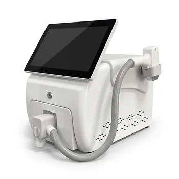
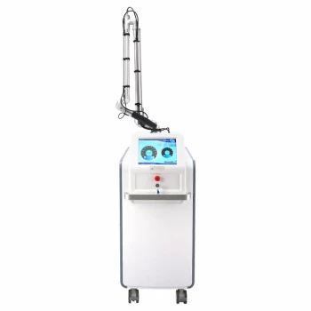
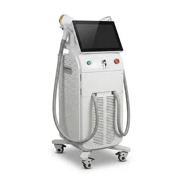
Ensure safe and effective vascular laser treatments with BELIS' advanced technology. Our professional-grade devices, including diode and Nd:YAG lasers, provide precise energy delivery with advanced cooling systems, minimizing the risk of non-specific thermal damage. For clinics and premium salons seeking superior safety and efficacy, contact us today to learn how BELIS can enhance your practice and patient outcomes.
Related Products
- Ultrasonic Cavitation Machine Lipo Laser Device
- Diode Laser SHR Trilaser Hair Removal Machine for Clinic Use
- 808nm Diode Laser Hair Removal Machine 755+808+1064nm Mixed Wavelength Professional Equipment
- 9D 7D HIFU Vaginal RF Lifting Treatment
- 808nm Diode Laser Hair Removal Machine and Equipment with Picolaser Arm
People Also Ask
- What are the clinical advantages and operational precautions associated with ultrasound cavitation technology in fat reduction equipment? Maximize Safety and Efficacy
- How do the physiological effects of acoustic cavitation and thermal stress on adipocytes explain the functional mechanism of non-invasive ultrasonic RF cavitation body sculpting equipment? | Key Insights
- Why are non-invasive RF skin tightening devices preferred over ablative laser resurfacing? Safe Operator Techniques
- How does patient age affect clinical outcomes and candidate selection for non-invasive radiofrequency (RF) skin tightening procedures? Age matters, but it's not the only criterion for success.
- How do non-invasive body sculpting technologies using RF and ultrasound address surgical fat reduction risks? Lower-risk body contouring with RF and ultrasound.