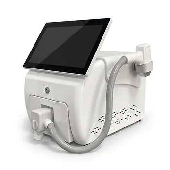
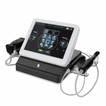
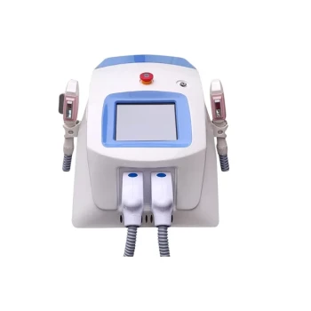
A history of post-injection granulomas or delayed inflammatory reactions requires a more cautious evaluation before any laser or energy-based treatment. The practitioner should determine whether inflammation is active, identify the injected material and treatment location, and assess tissue health with appropriate examination and imaging. Thermal or ablative energy should not be applied over active nodules, unresolved granulomas, acutely inflamed skin, compromised wounds, or areas containing unknown or permanent fillers.
The key decision is not simply whether the patient has had a granuloma, but whether residual foreign material or active inflammation remains in the treatment field. Resolve and characterize the inflammatory process first; if the filler composition or tissue status is uncertain, defer treatment or avoid that anatomical area.
Establish Whether the Reaction Is Active
Look for clinical signs of ongoing inflammation
Delayed reactions may present weeks after injection as hardened erythema, palpable nodules, tenderness, edema, or localized inflammatory infiltrates. These findings can represent delayed hypersensitivity, foreign-body granuloma, infection, or another inflammatory disorder.
Treatment should be postponed when nodules are enlarging, painful, warm, draining, erythematous, or otherwise clinically active. Applying heat to such tissue can intensify inflammatory signaling and potentially worsen the reaction.
Distinguish a stable history from unresolved disease
A remote, fully resolved reaction does not automatically prohibit every future energy-based treatment. However, the practitioner should establish when the reaction occurred, how it was treated, whether it recurred, and whether any nodules or induration remain.
A patient who reports intermittent swelling, recurrent redness, persistent firmness, or unexplained tenderness should not be treated as having a purely historical problem.
Exclude infection and other mimics
A presumed granuloma may instead be an infection, biofilm-associated process, cyst, scar nodule, or another lesion requiring different management. Suspicious, atypical, or worsening findings warrant appropriate medical assessment and, when indicated, referral for diagnostic imaging, laboratory evaluation, or tissue diagnosis.
Identify the Injected Material and Its Location
Obtain complete filler documentation
Review the product name, injection date, anatomical plane, volume, treatment provider, and any subsequent procedures. The practitioner should also document prior complications and whether the reaction followed a specific filler or a particular energy-based treatment.
This information matters because degradable hyaluronic-acid fillers and permanent or non-biodegradable materials do not carry identical clinical implications.
Treat unknown or permanent fillers as a major risk factor
Liquid silicone, acrylic hydrogels, and other non-biodegradable materials can remain in tissue and may produce unpredictable inflammatory responses. High-energy or ablative treatments over these areas may trigger a severe flare, sometimes described as a “burst granuloma,” and can complicate later diagnosis and management.
If the filler composition cannot be definitively established, energy treatment in that anatomical region should generally be avoided until the risk has been properly assessed.
Consider the treatment field, not only the injection history
A prior filler reaction elsewhere on the face is not equivalent to an active or permanent filler deposit directly beneath the proposed treatment area. Map previous injection sites against the planned laser or radiofrequency field.
When the relationship between the filler and the treatment zone is unclear, diagnostic imaging can help characterize tissue changes and guide a safer decision.
Use Examination and Imaging Before Treatment
Perform a structured skin and tissue assessment
Assess baseline erythema, edema, induration, nodularity, tenderness, pigmentation, scarring, and wound integrity. Compare the proposed treatment area with adjacent uninvolved skin when possible.
The assessment should also include current medications, allergies, autoimmune or inflammatory disease, infection risk, prior wound-healing problems, and any history of hypertrophic scars or keloids.
Use skin diagnostic imaging when appropriate
Pre-treatment imaging with suitable skin diagnostic devices can help identify underlying inflammation, residual nodules, fluid collections, or tissue abnormalities that are not obvious on casual inspection. Imaging does not replace clinical judgment or specialist evaluation, but it can reduce the risk of treating apparently normal skin over abnormal tissue.
Documenting baseline findings is also valuable if delayed swelling, erythema, or textural changes develop later.
Choose the Device and Timing Conservatively
Avoid thermal energy over active inflammatory tissue
Laser, radiofrequency, and other thermal systems should not be used directly over active granulomas, unresolved inflammatory nodules, or acutely inflamed skin. Additional heat may aggravate the local immune response and expand tissue injury.
The same principle applies to compromised wounds, active infection, and untreated inflammatory eruptions.
Separate inflammation control from scar remodeling
If the patient has both a prior inflammatory reaction and acne scarring, first stabilize the inflammatory process. Active acne should be adequately controlled before invasive resurfacing because treatment may worsen inflammation, reactivate deep cyst formation, or increase scarring.
Once the active phase has settled, a staged approach may be considered. Vascular-selective devices such as IPL or pulsed-dye laser may address persistent red scar borders, while fractional laser or RF micro-remodeling may later address textural changes.
Use lower-risk parameters when treatment is appropriate
For a patient with resolved inflammation and no contraindicating filler material, use conservative fluence, limited thermal overlap, and an appropriate fractional rather than aggressive non-fractionated approach when clinically suitable.
Patients with Fitzpatrick skin types IV–VI require particular caution because they have higher risks of post-inflammatory hyperpigmentation, hypertrophic scarring, and keloid formation. Pigment-prevention strategies and extended follow-up may be appropriate.
Review Broader Contraindications
Check medications and healing risk
Screen for isotretinoin use, photosensitizing medications, anticoagulants, impaired wound healing, active infection, and relevant systemic disease. Guidance on the interval after isotretinoin varies by procedure, device, and current evidence; the practitioner should follow the device manufacturer’s instructions and applicable clinical guidance rather than rely on a universal interval.
For invasive laser scar treatments, many protocols defer treatment for at least six months after isotretinoin, while some more conservative protocols use longer intervals. The decision should be individualized and documented.
Assess skin type and scar history
Ask about personal and family histories of hypertrophic scars and keloids, particularly before procedures that create significant thermal or mechanical injury. A previous injection granuloma is not the same as a keloid tendency, but both histories may indicate the need for heightened caution and realistic counseling.
Screen for device-specific contraindications
Depending on the technology, assess pregnancy or nursing, implanted electrical devices, epilepsy, active cancer or malignant lesions, photosensitizing drugs, and other manufacturer-listed contraindications. These considerations are especially important for radiofrequency, intense pulsed light, and high-powered laser systems.
Plan Appropriate Observation and Follow-Up
Explain that reactions can be delayed
Patients with a history of delayed inflammatory reactions should understand that complications may appear days or weeks after treatment rather than immediately. Consent should address possible swelling, erythema, pigment alteration, nodule recurrence, scarring, and the possibility that further treatment may be required.
Use staged treatment and test areas when justified
A small test area may help assess the immediate tissue response, particularly when skin type, filler history, or treatment parameters create uncertainty. A test spot cannot guarantee that delayed inflammation will not occur, so it should not replace filler identification, clinical evaluation, or appropriate deferral.
Extend follow-up for higher-risk patients
Arrange early review and maintain longer observation when the patient has darker skin, a significant scar history, prior granulomatous reactions, uncertain filler history, or treatment involving substantial thermal injury. Follow-up intervals of six to eight weeks may be more appropriate than routine shorter intervals when delayed hyperplastic or inflammatory responses are a concern.
Understanding the Trade-offs
Treating too early can worsen the original problem
The main risk is not merely transient redness. Energy applied over residual foreign material or active inflammation can produce a larger inflammatory flare, tissue damage, pigmentary change, or more difficult diagnostic and treatment course.
Deferral may be frustrating for the patient, but it is clinically preferable to converting a localized, manageable problem into a more extensive reaction.
Avoiding treatment indefinitely may also be unnecessary
A prior reaction does not automatically mean that all future laser or energy-based treatments are contraindicated. Once the reaction is clinically inactive, the material is known or appropriately assessed, and the treatment field is safe, a carefully selected and conservative procedure may be reasonable.
The decision should be based on current tissue status rather than the historical label alone.
Pain control introduces its own safety requirements
Painful procedures may involve topical anesthesia, injected local anesthetic, or sedation. These can cause complications such as respiratory depression, hypoxemia, swallowing difficulty, or hypotension.
Clinics should complete pre-treatment screening, monitor vital functions when indicated, observe patients after treatment, and maintain appropriate emergency protocols. Sedation should never be used to bypass uncertainty about whether the procedure itself is safe.
Financial pressure must not determine candidacy
Elective treatment should be declined or deferred when the risk of severe inflammation, scarring, infection, or pigmentary alteration is unacceptably high. The practitioner should explain the clinical reason and offer appropriate alternatives or referral rather than proceed for financial reasons.
How to Apply This to the Patient’s Situation
Use the following decision framework before scheduling treatment:
- If your primary focus is identifying active inflammation: Defer laser or energy treatment, document the findings, and arrange appropriate medical evaluation until nodules, erythema, tenderness, or other inflammatory signs have resolved.
- If your primary focus is determining filler-related risk: Obtain product and injection records, map the treatment field, and use diagnostic imaging or specialist assessment when the material or residual deposits are uncertain.
- If your primary focus is treating acne scarring: Control active acne first, then consider staged vascular and fractional remodeling treatments only after the inflammatory phase has stabilized.
- If your primary focus is minimizing pigment or scar complications: Account for Fitzpatrick skin type, personal and family scar history, use conservative parameters, and plan extended follow-up.
- If your primary focus is procedural safety: Review medications, isotretinoin history, systemic contraindications, anesthesia risks, and device-specific warnings before consenting the patient.
The safest approach is to treat the patient’s current tissue status—not merely the cosmetic indication—before deciding whether energy-based treatment is appropriate.
Summary Table:
| Consideration | Key Points |
|---|---|
| Active Inflammation | Defer treatment if nodules, erythema, tenderness, or drainage present. |
| Filler History | Document product, date, location; unknown or permanent fillers increase risk. |
| Imaging | Use skin diagnostic tools to identify hidden inflammation or nodules. |
| Device Selection | Avoid thermal energy over active lesions; use conservative parameters. |
| Medication Review | Screen for isotretinoin, photosensitizers, anticoagulants. |
| Follow-up | Extend to 6-8 weeks for high-risk patients; monitor for delayed reactions. |
Ensure patient safety and optimal outcomes with BELIS's advanced aesthetic devices, designed for clinics and premium salons. Discover our range of laser, IPL, and body contouring systems that meet rigorous safety standards. Contact us today for expert guidance and support.
Related Products
- Professional Face and Vaginal 7D HIFU System for HIFU Clinic Treatments
- 22D HIFU Machine Device Facial Machine
- Trilaser Diode Hair Removal Machine for Beauty Clinic Use
- Tri Laser Diode Hair Removal Machine Professional Beauty Equipment
- Cryolipolysis Fat Freezing Machine and Ultrasonic Cavitation Device
People Also Ask
- How does focused ultrasound (HIFU) technology produce non-invasive tissue tightening in deep dermal layers? Understand the exact mechanism and discover expert treatment insights.
- What is the mechanism of action of High-Intensity Focused Ultrasound (HIFU) devices in noninvasive body sculpting, and how is surrounding tissue protected?
- How does the safety profile of energy-based skin tightening equipment, such as HIFU and microneedle RF, compare to injectable fillers regarding vascular occlusion risks?
- How do HIFU and Microneedle RF compare to Botulinum Toxin for upper facial complications?
- How does non-invasive submental tightening with HIFU align with conservative volume preservation to avoid contour deformities?