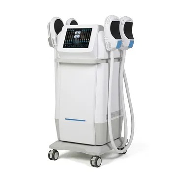
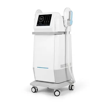
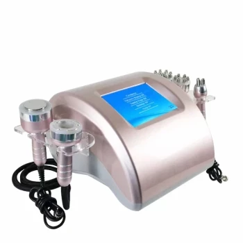
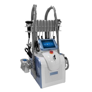
Subcutaneous white adipose tissue (WAT) determines both what non-invasive body sculpting can treat and how it should be applied. Its depth, lobular organization, fibrous septa, vascularity, and relationship to the skin guide patient selection, applicator placement, energy or cooling settings, and treatment expectations. Cryolipolysis primarily targets pinchable adipose tissue through controlled cooling; radiofrequency and therapeutic ultrasound use thermal or acoustic effects; high-intensity electromagnetic systems primarily stimulate muscle and may complement, rather than directly replace, fat-reduction technologies.
The key principle is anatomical matching: non-invasive equipment is most predictable when a clinician identifies a localized, accessible subcutaneous fat depot and selects a modality whose physical mechanism matches the tissue depth, thickness, mobility, and treatment goal.
Why WAT Structure Matters in Body Sculpting
WAT is a localized energy reservoir
Subcutaneous WAT consists mainly of mature adipocytes containing a large lipid droplet. These cells are organized into lobules within a connective-tissue matrix and supported by small blood vessels, nerves, and lymphatic structures.
This makes WAT different from visceral fat, which surrounds internal organs and cannot be safely reached by external body-contouring applicators. Non-invasive body sculpting is therefore designed for localized subcutaneous adiposity, not general weight reduction or visceral-fat treatment.
Adipocyte size influences visible contour
Changes in WAT volume mainly reflect changes in adipocyte size, although cell number and depot architecture also contribute. A patient may have a normal overall body weight but still possess a discrete abdominal, flank, thigh, or submental fat pocket.
This is why treatment assessment should focus on localized contour anatomy, not BMI alone. The practical question is whether a measurable, accessible fat layer exists that can be treated without compromising the skin or underlying structures.
The tissue is organized into layers
Subcutaneous fat is commonly described as superficial and deep compartments, separated in some regions by fascial structures such as Scarpa’s fascia. The superficial layer is more constrained by fibrous septa, while the deeper layer is generally looser.
Layering affects applicator contact, cooling or heating distribution, tissue penetration, and the likelihood of treating the intended depot. Clinicians should assess the pinchable fat thickness and its location, rather than assuming that all visible fullness represents the same tissue compartment.
How Anatomy Guides Modality Selection
Cryolipolysis requires an accessible fat fold
Cryolipolysis uses controlled cooling to create injury within susceptible adipocytes while aiming to preserve the overlying skin and surrounding tissues. It is best suited to localized, pinchable subcutaneous fat that can be drawn into the applicator.
The thickness, mobility, and shape of the fat fold influence applicator selection and positioning. Areas with insufficient tissue capture, marked fibrosis, or unfavorable anatomy may produce less predictable results or increase the risk of an uneven contour.
Radiofrequency is primarily a thermal modality
Radiofrequency devices deliver electromagnetic energy that produces tissue heating. Depending on the device and protocol, the intended effects may include temporary tissue warming, collagen remodeling, skin tightening, and an adjunctive contribution to contour improvement.
RF should not automatically be described as selectively destroying adipocytes. Its clinical effect depends on the device’s frequency, electrode configuration, energy delivery, temperature monitoring, treatment duration, and regulatory indications.
Ultrasound cavitation should be distinguished from RF
“Cavitation” generally refers to an acoustic ultrasound mechanism, not radiofrequency itself. Ultrasound may generate mechanical effects in tissue, but the exact biological response depends on the device design, operating parameters, tissue composition, and treatment protocol.
Clinicians should therefore distinguish RF heating, therapeutic ultrasound, and other energy modalities when explaining treatment mechanisms. Using precise terminology prevents unrealistic expectations and improves protocol selection.
Electromagnetic systems primarily address muscle
High-intensity electromagnetic body-contouring systems induce repeated muscle contractions. Their principal target is neuromuscular tissue, and their role is generally to improve muscle conditioning or definition rather than directly remove a subcutaneous fat pocket in the same manner as cryolipolysis.
They may be useful when the patient’s goal includes abdominal or gluteal muscle development. They should not be presented as interchangeable with a device specifically intended for localized adipose reduction.
What the Clinician Must Assess Before Treatment
Measure fat thickness and tissue mobility
A clinical examination should identify whether the fullness is soft and mobile, dense and fibrotic, thin and superficial, or primarily related to skin laxity. Pinch testing, visual assessment, palpation, and standardized photographs help establish whether the target is suitable for an external applicator.
Very thin fat layers provide limited treatment volume and may increase the risk of an irregular appearance. Conversely, substantial but anatomically accessible fat may permit more reliable applicator coupling or tissue capture.
Evaluate the overlying skin
Skin elasticity determines how well the surface may conform after a modest reduction in underlying volume. Good or moderate elasticity generally supports a smoother visual result.
When laxity is the dominant problem, fat reduction alone may worsen apparent looseness. RF-based skin-heating protocols may offer some remodeling potential, but severe laxity may require a surgical discussion rather than repeated non-invasive fat treatments.
Identify fibrous structures and adherence zones
Fibrous septa organize fat lobules and connect the skin to deeper tissues. In areas such as the infragluteal fold and mid-medial thigh, strong tissue attachments help define normal contour and should not be treated as simple excess fat.
Non-invasive methods generally preserve tissue continuity better than surgical excision, but poor applicator placement or excessive treatment can still create asymmetry. Anatomical landmarks must guide treatment boundaries.
Differentiate fat from cellulite
Excess adiposity is an increase in volume of otherwise relatively normal adipose tissue. Cellulite involves a broader structural pattern, including fibrous septal tethering, skin surface changes, and focal fat herniation.
A fat-reduction device may reduce volume without correcting the septal attachments that produce dimpling. Patients seeking cellulite improvement may require a different tissue-remodeling strategy or a combination approach.
Exclude visceral fullness
External applicators cannot selectively treat fat located inside the abdominal cavity. A protruding abdomen may reflect visceral adiposity, abdominal wall structure, posture, bloating, or skin laxity rather than a treatable subcutaneous pocket.
The patient must understand that body contouring is a localized aesthetic intervention, not a substitute for weight management or treatment of metabolic disease.
How Tissue Physiology Influences Treatment Planning
The microvascular network matters
Subcutaneous WAT contains a capillary network and connective matrix that support tissue viability and transport. The goal of a well-designed protocol is to affect the intended adipose target while limiting injury to skin, nerves, vessels, and connective tissue.
This is why manufacturer-defined applicator placement, contact conditions, temperature limits, and treatment durations matter. Increasing energy or extending treatment time beyond validated parameters does not automatically improve results.
Tissue consistency affects energy delivery
Soft, mobile fat and dense, fibrous fat do not respond identically to external treatment. Fibrosis can alter coupling, mechanical transmission, and the distribution of thermal or acoustic energy.
A previously treated area may also have different tissue compliance from untreated WAT. Treatment history should therefore be included in the assessment rather than relying only on the visible contour.
Vascularity affects safety considerations
The target is not an isolated mass of lipid. Blood vessels, nerves, fascia, and skin are interwoven with or adjacent to the fat layer.
Protocols must account for the possibility of thermal injury, cold-related injury, pain, sensory changes, bruising, or contour irregularity. Device-specific contraindications and safety screening remain essential because tissue anatomy alone cannot establish clinical eligibility.
Treatment effects are not immediate volume evacuation
Non-invasive methods do not remove adipose tissue immediately as liposuction does. Depending on the modality, the intended process may involve controlled adipocyte injury, gradual clearance of cellular material, tissue remodeling, or changes in muscle tone.
Patients should expect a progressive and often modest contour change rather than an instant surgical-style reduction. Follow-up photography and standardized measurements are more reliable than day-to-day visual inspection.
Understanding the Trade-offs
Non-invasive treatment is not equivalent to surgery
Non-invasive procedures avoid incisions, tissue aspiration, and anesthesia, which can reduce procedural burden. They also generally provide less dramatic and less controllable volume reduction than surgical excision or liposuction.
The appropriate comparison is therefore not “which method is universally better,” but which method matches the amount of tissue change required and the patient’s risk tolerance.
More energy is not necessarily better
Excessive cooling, heating, acoustic exposure, or electromagnetic intensity can increase adverse effects without guaranteeing better contouring. Tissue safety depends on controlled exposure and accurate application, not on maximizing the device output.
Clinicians should use validated protocols and document the treated area, applicator, settings, contact conditions, and patient response.
Skin laxity can limit the result
Reducing a fat pocket beneath poorly elastic skin may leave the patient dissatisfied, even if adipose volume decreases. The visible problem may be driven more by skin redundancy than by fat thickness.
A candid consultation should distinguish fat reduction, skin tightening, muscle definition, and cellulite improvement as separate objectives.
Cellulite may persist
Because cellulite involves septal tethering and surface architecture, reducing adipocyte volume may not eliminate dimpling. Treating the wrong structural problem is a common source of dissatisfaction.
Clinical assessment should identify whether the patient’s priority is a smaller fat pocket, smoother skin, improved laxity, or a combination of these outcomes.
Results vary across body regions and patients
WAT thickness, septal organization, dermal thickness, collagen elasticity, and prior treatment history differ between individuals and anatomical sites. These variables create meaningful variation in response.
A standardized protocol can improve consistency, but it cannot eliminate biological variability. Outcome claims should remain proportional to the treatment area and the device’s established indication.
Applying WAT Physiology in the Clinic
A practical assessment should connect the patient’s goal to the underlying tissue before selecting equipment.
- If your primary focus is localized fat reduction: Confirm a discrete, pinchable subcutaneous depot and choose a modality and applicator validated for that tissue thickness and body region.
- If your primary focus is skin laxity: Assess dermal elasticity and consider whether a controlled thermal remodeling approach is appropriate; severe laxity may require surgical evaluation.
- If your primary focus is muscle definition: Consider a high-intensity electromagnetic system as a muscle-focused treatment rather than representing it as a direct substitute for adipose-targeting technology.
- If your primary focus is cellulite: Diagnose fibrous septal tethering and skin-surface architecture first, because adipocyte volume reduction alone may not correct the underlying dimpling.
- If your primary focus is abdominal fullness: Distinguish accessible subcutaneous fat from visceral adiposity, abdominal wall factors, and generalized weight gain before promising contour improvement.
- If your primary focus is safety and predictability: Match treatment depth, applicator geometry, energy or cooling parameters, and treatment boundaries to the patient’s anatomy and the device’s validated instructions.
The most reliable body-sculpting decisions begin with identifying the tissue—not merely the body area—and matching the technology to its structure and the patient’s actual goal.
Summary Table:
| Tissue Feature | Clinical Relevance | Assessment Method |
|---|---|---|
| Thickness | Determines if fat is pinchable and suitable for applicator-based devices | Pinch test, calipers |
| Mobility | Influences applicator contact and treatment efficacy | Palpation, pinch test |
| Fibrosis | Can affect energy/cooling distribution and treatment results | Palpation, history |
| Vascularity | Affects safety and risk of adverse effects | Medical history, clinical judgment |
| Skin Elasticity | Impacts final contour and need for skin tightening | Visual, pinch test |
| Compartment | Superficial vs. deep affects applicator selection | Ultrasound, clinical assessment |
Ready to enhance your practice with evidence-based body sculpting? Contact BELIS today to discover our advanced aesthetic devices, including cryolipolysis, RF, HIFU, and EMS systems, tailored for clinics and premium salons. Our experts will help you select the right technology to match your patients' anatomy and goals. Contact us now to discuss your needs and learn about our OEM/ODM support and certification.
Related Products
- EMSlim Neo Nova Body Sculpting EMS Sculpting Machine
- EMS Body Sculpting Slimming Machine EMSlim Body Slimming Machine
- EMS Body Sculpting Machine for Body Slimming and Sculpting
- EMSlim RG Laser Body Sculpting and Slimming Machine
- EMSlim Body Slimming Machine for Muscle Building and Beauty
People Also Ask
- How do RF body sculpting devices target subcutaneous fat & tighten skin?
- What is the biological mechanism of action by which radiofrequency (RF) energy achieves body sculpting and cellulite reduction? Discover how controlled heating remodels collagen, influences fat, and smooths skin.
- What anatomical mechanisms cause lower face and submental contour loss, and how do body sculpting and RF technologies address jowls and neck laxity? Discover the key causes and non-surgical solutions.
- How can aesthetic practitioners accurately differentiate between muscular hypertrophy and subcutaneous fat accumulation when evaluating patients for calf or lower face contouring using body sculpting equipment?
- How does radiofrequency (RF) operating frequency influence tissue selectivity and therapeutic outcomes in non-invasive RF body sculpting systems?