The key is to identify the anatomical layer before choosing a device. To distinguish muscular hypertrophy from subcutaneous fat, assess the area both at rest and during selective muscle contraction, then confirm the findings with objective imaging—preferably high-frequency ultrasound. Tiptoe standing can expose gastrocnemius enlargement in the calf, while jaw clenching can reveal masseter hypertrophy in the lower face; soft, mobile tissue that remains outside the contracting muscle is more consistent with subcutaneous fat.
Do not treat “volume” as a diagnosis. Determine whether the excess comes from muscle, fat, bone, glandular tissue, fluid, or a pathological mass before selecting body-sculpting equipment.
Start With a Layer-by-Layer Examination
Examine the patient at rest
Document the contour from multiple angles and compare both sides for symmetry, borders, skin quality, and visible muscle definition.
Palpation should assess softness, mobility, firmness, tenderness, and adherence to deeper structures. Fat is usually pliable and mobile relative to the underlying fascia, whereas a contracted muscle becomes firm and more prominent.
Repeat the examination during contraction
Dynamic examination is particularly useful because muscle volume changes with activation while subcutaneous fat does not.
For the calf, ask the patient to perform a controlled heel raise or stand on tiptoe. A prominent, firm bulge that increases during plantar flexion suggests gastrocnemius involvement; deeper fullness may involve the soleus and is more difficult to assess by inspection alone.
For the lower face, ask the patient to clench the teeth briefly while the examiner palpates the masseter. A visible or palpable increase in the lateral jaw angle supports masseter hypertrophy, although clenching should be brief and avoided in patients with pain or temporomandibular disorders.
Compare contraction and relaxation
A useful clinical comparison is:
- Muscular predominance: firm enlargement, stronger definition during contraction, and a clear relationship to the muscle’s anatomical boundaries.
- Fat predominance: soft or compressible fullness that changes little with contraction and can often be pinched independently of the muscle.
- Mixed anatomy: a firm deep component combined with a superficial pinchable layer.
This distinction matters because reducing superficial fat will not substantially narrow a contour created mainly by hypertrophied muscle.
Use Imaging to Confirm the Tissue Layer
High-frequency ultrasound is the practical first-line tool
Ultrasound can help measure skin, subcutaneous fat, fascia, and muscle thickness along standardized anatomical reference points.
For reliable comparisons, use consistent patient positioning, probe orientation, measurement landmarks, and minimal probe pressure. Excessive compression can artificially reduce the apparent thickness of soft tissue.
Ultrasound is especially helpful when palpation cannot determine whether fullness lies above or within the muscle. It can also identify unexpected cystic, vascular, glandular, or solid lesions that should not be treated with routine body-contouring equipment.
Standardize calf measurements
Calf assessment should document the level of measurement, limb position, muscle state, and side-to-side differences.
Measure the tissue with the patient relaxed and, when clinically useful, repeat the assessment during contraction. The scan should distinguish the superficial subcutaneous layer from the gastrocnemius and deeper soleus compartments.
Standardize lower-face measurements
Lower-face width should not automatically be attributed to either fat or masseter muscle. Evaluate at least four contributors:
- Mandibular bone shape
- Masseter thickness
- Subcutaneous and buccal fat
- Parotid or other glandular enlargement
Ultrasound can help assess the superficial fat layer and masseter thickness. If the findings are atypical, asymmetric, painful, rapidly progressive, or poorly defined, medical evaluation and more advanced imaging may be appropriate.
Interpret the Clinical Findings Correctly
Fat is a superficial volume problem
Subcutaneous fat is located beneath the skin and above the muscle fascia. It generally produces a soft, compressible layer that can be measured and, in suitable patients, may be addressed with a fat-reduction modality.
The presence of fat does not automatically establish treatment suitability. The clinician must also assess skin elasticity, the amount of tissue available for treatment, and whether the expected improvement is clinically meaningful.
Muscle is a contractile volume problem
Muscular hypertrophy may result from activity, occupational loading, exercise, gait mechanics, or other causes. It is identified by increased muscle thickness and a clear change in firmness or contour during activation.
A device designed to reduce superficial adipose tissue is not a substitute for a treatment plan directed at muscle when muscle is the primary source of fullness.
Fluid and disease can mimic either condition
Swelling should be assessed for pitting, non-pitting character, warmth, tenderness, vascular changes, and time course. Unilateral or rapidly developing calf enlargement warrants appropriate medical assessment rather than elective contouring.
A firm mass, unexplained asymmetry, pain, skin changes, or a lesion that is not clearly normal subcutaneous fat should be investigated before energy-based treatment. Non-invasive body-sculpting devices are intended for appropriate healthy tissue, not unexamined tumors, inflammatory lesions, or encapsulated masses.
Match the Treatment to the Diagnosis
When subcutaneous fat is the dominant finding
Fat-reduction technologies may be considered when imaging confirms an adequate superficial adipose layer and the patient has realistic expectations.
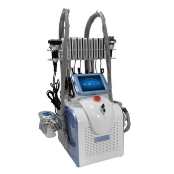
Depending on the device and its validated indication, options may include cryolipolysis, radiofrequency-based treatment, or focused ultrasound. The specific mechanism, treatment depth, contraindications, and regulatory authorization must be verified for the device and anatomical site.
These treatments affect the superficial fat compartment. They do not reliably correct bony width, substantial muscle hypertrophy, visceral fat, glandular enlargement, or significant skin laxity.
When muscle hypertrophy is dominant
If the calf or lower-face contour is primarily muscular, superficial fat reduction is likely to produce little change.
The practitioner should explain that the treatment target is different and consider referral or a clinically appropriate muscle-directed approach within the practitioner’s scope, training, and local regulations. The diagnosis should be documented rather than inferred from the patient’s request for “fat reduction.”
When the findings are mixed
Many patients have both muscle and fat contributing to the contour. In these cases, quantify the relative contribution before proposing staged treatment.
Treating only the superficial layer may create an underwhelming result, while excessive reduction around the lower face can produce an unnecessarily hollow appearance. A conservative plan is preferable when the anatomical contribution is uncertain.
When skin laxity is significant
Reducing fat in loose skin can improve volume but may leave residual laxity or make laxity more apparent.
Skin quality should therefore be assessed separately from fat thickness. A plan may require a skin-tightening strategy, no treatment, or referral rather than fat reduction alone.
Understand the Trade-offs
Palpation is useful but not definitive
Dynamic palpation is fast and clinically valuable, but it is operator-dependent. It can be misleading in patients with edema, dense fibrous tissue, substantial body mass, or mixed muscle-fat anatomy.
Imaging improves objectivity, but it does not replace a complete clinical examination or appropriate medical referral when findings are atypical.
Device labels do not establish tissue diagnosis
Terms such as RF, cavitation, HIFU, cryolipolysis, and EMS describe technologies, not diagnoses.
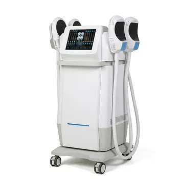
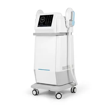
For example, electromagnetic muscle-stimulation systems primarily produce controlled muscle contractions and should not be presented as equivalent to a device that directly reduces subcutaneous fat. Treatment claims must match the device’s actual mechanism, evidence, and authorized indication.
The calf is not automatically a suitable fat-treatment area
Some calves contain little pinchable subcutaneous fat and are shaped mainly by the gastrocnemius, soleus, tendon, and bone.
Treating an insufficient fat layer can produce minimal benefit and may increase dissatisfaction. Practitioners should be willing to decline treatment when the target tissue is absent or the expected contour change is too small.
Lower-face contouring has a narrow margin for error
The lower face contains closely related aesthetic structures, including muscle, fat, glands, and bone.
Over-reducing superficial fat can create hollowing, while misidentifying parotid or masseter enlargement can lead to an ineffective or inappropriate treatment. Conservative treatment planning and standardized photography are essential.
Cellulite is not the same as excess fat
Cellulite involves changes in the relationship between fat, skin, and fibrous septa. A fat-volume reduction approach may not correct the dimpling or dermal tethering that defines cellulite.
The practitioner should identify whether the patient’s concern is localized volume, surface irregularity, skin laxity, or a combination of these conditions.
How to Apply This to Your Assessment
Use a consistent protocol before treating either the calf or lower face:
- If your primary focus is diagnostic accuracy: Examine the area at rest and during selective contraction, then confirm the suspected layer with standardized high-frequency ultrasound.
- If your primary focus is calf contouring: Separate superficial fat from gastrocnemius and soleus volume, and refer unilateral, painful, rapidly changing, or unexplained swelling for medical evaluation.
- If your primary focus is lower-face contouring: Assess mandibular structure, masseter thickness, superficial or buccal fat, and parotid size before selecting a modality.
- If your primary focus is fat reduction: Treat only when an adequate, accessible subcutaneous fat layer is documented and the device is appropriate for that tissue and anatomical site.
- If your primary focus is patient safety: Exclude unexamined masses, significant fluid-dominant swelling, active disease, and unrealistic expectations before treatment.
- If your primary focus is outcome quality: Account separately for muscle, fat, skin laxity, and bone so that reducing one layer does not create an imbalanced or hollowed contour.
Accurate contouring begins with identifying the tissue responsible for the volume—not with choosing a device.
Summary Table:
| Feature | Muscular Hypertrophy | Subcutaneous Fat Accumulation |
|---|---|---|
| Palpation at rest | Firm, well-defined | Soft, compressible, mobile |
| Response to contraction | Increases in size and firmness | Minimal change |
| Ultrasound appearance | Increased muscle thickness | Increased subcutaneous fat layer |
| Treatment approach | Muscle-directed (e.g., EMS) | Fat reduction (e.g., cryolipolysis) |
Ensure accurate diagnosis and optimal outcomes for your patients. Contact BELIS today to learn about our advanced diagnostic tools and body sculpting equipment tailored for clinics and premium salons. Our portfolio includes HIFU, cryolipolysis, and more. Contact us now to elevate your practice.
Related Products
- EMSlim Neo Nova Body Sculpting EMS Sculpting Machine
- EMS Body Sculpting Machine for Body Slimming and Sculpting
- EMS Body Sculpting Slimming Machine EMSlim Body Slimming Machine
- EMSlim Body Sculpting Machine EMS Body Slimming Machine
- EMSlim Body Slimming Machine for Muscle Building and Beauty
People Also Ask
- What anatomical mechanisms cause lower face and submental contour loss, and how do body sculpting and RF technologies address jowls and neck laxity? Discover the key causes and non-surgical solutions.
- How do the mid-infrared transmission spectra of fatty tissue and lipids assist in understanding selective targeting for body sculpting devices like cryolipolysis and RF cavitation? Mid-infrared spectra reveal lipid-specific markers, aiding tissue characterization.
- How should aesthetic clinics evaluate post-massive weight loss patients using tissue assessment techniques like the pinch test to determine suitability for non-invasive body sculpting devices versus surgical tissue excision? A Comprehensive Guide
- What are the VTE safety advantages of non-invasive body sculpting over surgery? Lower thromboembolic risk without general anesthesia
- What common soft tissue complications follow surgical fat extraction, and how do non-invasive body sculpting modalities eliminate these risks? Discover safer alternatives for your patients