Mid-infrared transmission spectra help identify what makes adipose tissue chemically distinct, but they do not directly cause or independently control selective body sculpting. Fatty tissue and purified lipids show strong C–H stretching bands around 2850–2920 cm⁻¹ and an ester carbonyl feature near 1740 cm⁻¹, while muscle and other protein-rich tissues show stronger amide bands. These signatures help researchers understand tissue composition, validate models, and design energy-delivery strategies that preferentially affect subcutaneous fat while limiting exposure to skin, nerves, vessels, and muscle.
Core takeaway: Mid-infrared spectra provide a molecular fingerprint of lipid-rich tissue. They support the rationale for selective treatment, but cryolipolysis depends mainly on differential cold sensitivity and RF-based treatments depend mainly on electrical, thermal, and anatomical properties—not on mid-infrared absorption alone.
What the Spectra Reveal About Adipose Tissue
Lipid-specific C–H absorption bands
The bands near 2850–2920 cm⁻¹ arise primarily from C–H stretching vibrations in CH₂ and CH₃ groups, which are abundant in triglycerides and other lipid molecules.
Their prominence confirms that adipose tissue contains a high concentration of hydrocarbon-rich lipid structures compared with many surrounding tissues.
The ester carbonyl signature
The sharp feature near 1740 cm⁻¹ is associated with the ester carbonyl group found in triglycerides and other lipid esters.
Together with the C–H bands, this feature provides a useful spectroscopic indication that the sampled tissue is lipid-rich rather than predominantly protein-based.
Contrast with muscle and connective tissue
Muscle contains substantial protein and water, so its spectrum includes more prominent amide-related bands, particularly those associated with peptide bonds.
This contrast helps distinguish adipose tissue from muscle, dermal tissue, and other structures during laboratory characterization or tissue-imaging research.
How Spectral Differences Relate to Selective Targeting
Spectra establish a chemical basis for tissue discrimination
A transmission spectrum is effectively a molecular fingerprint. If two tissues absorb infrared energy differently, researchers can use that information to identify tissue composition and estimate where lipid-rich layers are located.
This is valuable for developing optical measurement systems, validating tissue models, and assessing whether an energy-based device is interacting with the intended anatomical layer.
Spectra are not the same as treatment selectivity
The presence of a lipid absorption peak does not mean that a cryolipolysis or RF device selectively targets fat through mid-infrared absorption.
Treatment selectivity depends on the relationship between energy delivery, tissue composition, heat or cooling transfer, exposure time, tissue depth, and biological response.
The practical role is usually indirect
In many systems, spectroscopy supports device development rather than serving as the treatment mechanism. It can help characterize tissue layers, test applicator assumptions, and identify the boundary between adipose tissue and structures that require protection.
This distinction is important because a spectrometer may identify fat molecularly, while a body sculpting applicator may use cooling, radiofrequency, ultrasound, or another physical mechanism.
How This Applies to Cryolipolysis
Cryolipolysis relies on differential cold sensitivity
Cryolipolysis applies controlled cooling to a localized tissue volume. Adipocytes are more vulnerable to the treatment’s cooling conditions than the overlying skin and several surrounding tissue structures.
The resulting cellular injury initiates a biological clearance process in which damaged fat cells are gradually removed over subsequent weeks to months.
Lipid chemistry helps explain the vulnerability
The lipid-rich composition identified by the infrared spectrum is relevant because adipocytes contain large intracellular lipid stores. Lipid behavior under cooling contributes to the different thermal response of fat compared with water-rich or protein-rich tissues.
However, selective cryolipolysis should not be described as a simple consequence of a single lipid freezing point. The outcome also depends on cooling magnitude, exposure duration, tissue geometry, perfusion, insulation, and the biological tolerance of neighboring tissues.
Spectroscopy can support treatment modeling
Mid-infrared data can help researchers distinguish adipose from muscle or skin when building tissue models. Those models can then be used to study how cooling propagates through a layered body region and how much protection is needed for the skin surface.
The spectrum therefore supports understanding and verification, while the actual clinical selectivity comes from controlled thermal management and tissue-specific biological response.
How This Applies to RF-Based Body Sculpting
RF selectivity is primarily electrical and thermal
Radiofrequency systems deliver alternating electrical fields that generate heat through tissue resistance and molecular charge movement. The resulting temperature distribution depends on conductivity, impedance, perfusion, geometry, electrode or applicator design, and feedback control.
These are not directly represented by mid-infrared absorption peaks.
Adipose tissue can respond differently to RF energy
Subcutaneous fat and surrounding tissues have different electrical and thermal properties. Those differences can influence where heat is generated, how quickly it spreads, and how efficiently it is removed by blood flow.
A device may exploit those contrasts to heat a target layer while controlling surface temperature and limiting exposure to deeper or adjacent structures.
“RF cavitation” requires technical clarification
Radiofrequency and cavitation are different modalities. RF produces heating through electrical energy, whereas cavitation generally refers to acoustic effects associated with ultrasound.
Some commercial systems combine RF and ultrasound and may market them together, but their selectivity should be analyzed separately rather than attributed to one shared infrared mechanism.
What Mid-Infrared Data Can Assist With
Mapping tissue composition
Spectral signatures can help identify whether a measured region is predominantly lipid, protein, or water-rich tissue.
This information is useful when estimating the location and composition of a subcutaneous fat layer before or during device development.
Validating tissue phantoms and simulations
Researchers can compare the infrared spectrum of a laboratory tissue phantom with that of real adipose tissue. A close match supports the use of the phantom for studying energy deposition, thermal behavior, or applicator geometry.
Spectral characterization is especially useful when a model must represent fat and muscle as chemically different materials.
Supporting depth and boundary assessment
Layer-specific optical measurements may help distinguish adipose tissue from dermal or muscular regions. This can assist in determining whether an applicator is likely to affect the intended layer or whether energy may extend beyond it.
The usefulness depends on the measurement geometry, penetration depth, scattering, and whether the device actually operates in a compatible optical wavelength range.
Understanding the Trade-offs
Mid-infrared penetration is limited
Mid-infrared wavelengths are strongly absorbed by water and other tissue constituents and generally have limited penetration in biological tissue. A transmission spectrum obtained from a thin sample should not automatically be interpreted as a direct map of deep fat in an intact body.
For deep subcutaneous targeting, other imaging or monitoring methods may be more practical.
Molecular selectivity does not guarantee anatomical selectivity
Even if adipose tissue has a distinctive spectrum, energy can spread beyond the intended layer. Skin thickness, fat depth, blood flow, applicator contact, and patient anatomy all affect treatment exposure.
Chemical identification must therefore be combined with thermal, electrical, and anatomical controls.
RF terminology and claims can be misleading
RF devices do not selectively heat fat simply because fat has a lipid spectrum. Their behavior depends on field distribution and tissue electrical properties, which can vary substantially between individuals and treatment regions.
Claims about heating “only adipocytes” or completely sparing surrounding structures are technically overstated. Real systems manage risk through controlled energy delivery, surface cooling, monitoring, and treatment limits.
Cryolipolysis is not a purely chemical process
The lipid peaks explain why adipose tissue is compositionally distinctive, but treatment response also depends on cooling conditions and biological clearance. Vascular response, tissue thickness, applicator fit, and individual healing can affect results and adverse-event risk.
Spectral information cannot by itself predict the amount of fat reduction or guarantee a particular clinical outcome.
How to Apply This to Device Evaluation
Mid-infrared spectra are most useful when treated as supporting evidence, not as a complete explanation of device performance.
- If your primary focus is tissue identification: Use the C–H and ester carbonyl bands to distinguish lipid-rich adipose tissue from protein-rich muscle, while accounting for water absorption and measurement limitations.
- If your primary focus is cryolipolysis: Use spectral composition to inform tissue models, but evaluate selectivity through cooling profiles, fat depth, surface protection, and biological response.
- If your primary focus is RF treatment: Prioritize electrical conductivity, impedance, thermal diffusion, perfusion, field geometry, and temperature monitoring rather than mid-infrared peaks.
- If your primary focus is combined RF and cavitation equipment: Analyze RF heating and ultrasound cavitation as separate mechanisms with separate safety and targeting assumptions.
Used in the right context, mid-infrared spectroscopy clarifies what adipose tissue is made of, while device physics determines how selectively that tissue can be treated.
Summary Table:
| Aspect | Key Information |
|---|---|
| Lipid-specific absorption bands | C-H stretching around 2850-2920 cm⁻¹; ester carbonyl near 1740 cm⁻¹ |
| Contrast with other tissues | Muscle shows more amide bands, distinguishing adipose from protein-rich tissues |
| Role in selective targeting | Supports tissue modeling and validation, but selectivity depends on energy delivery and thermal/electrical properties |
| Application in cryolipolysis | Uses differential cold sensitivity; spectral data helps model tissue layers |
| Application in RF-based devices | Selectivity based on electrical and thermal properties, not mid-infrared absorption |
Optimize Your Body Sculpting Practice with BELIS
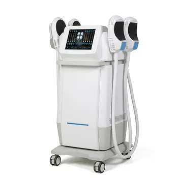
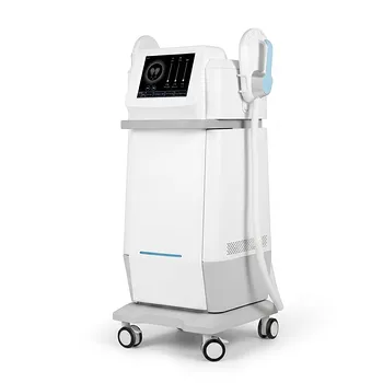
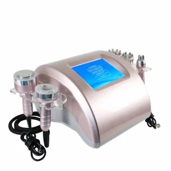
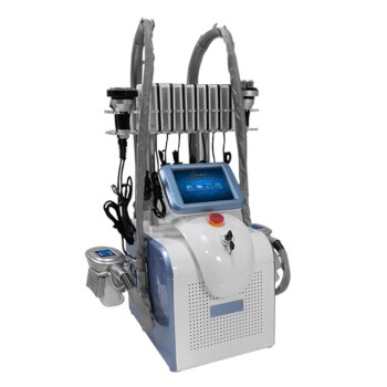
At BELIS, we provide advanced aesthetic equipment including cryolipolysis, RF cavitation, and a full range of laser and energy-based systems. Our devices are designed for clinics and premium salons, prioritizing safety, efficacy, and patient satisfaction. To schedule a consultation and discover how our technologies can enhance your offerings, contact us today.
Related Products
- EMS Body Sculpting Machine for Body Slimming and Sculpting
- EMSlim Neo Nova Body Sculpting EMS Sculpting Machine
- EMSlim Body Slimming Machine for Muscle Building and Beauty
- EMS Body Sculpting Slimming Machine EMSlim Body Slimming Machine
- EMSlim Body Sculpting Machine EMS Body Slimming Machine
People Also Ask
- Does EMS sculpting really work? Achieve Clinically-Proven Body Contouring
- Why is it important for practitioners to avoid over-promising body sculpting results? Ensure Trust and Clinic Success.
- What are the technical advantages of professional body sculpting? Non-Invasive vs. Invasive Liposuction Explained
- Does EMS actually work for weight loss? A Realistic Look at Muscle Building and Metabolism
- Why are non-invasive body sculpting devices preferred over surgery? Safe, Precise Fat Reduction for Premium Clinics