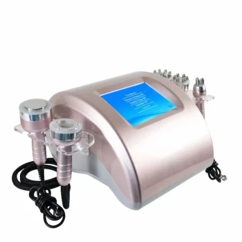
Real-time ultrasound visualization gives MFU/HIFU treatment a direct view of the tissue it is about to heat. It allows the clinician to assess skin thickness and tissue planes, select an appropriate focal depth, and confirm that thermal energy lines are positioned in the intended dermal or subcutaneous layer. Its primary clinical value is therefore greater treatment precision and safety, rather than simply greater energy output or automatically better tightening results.
Real-time imaging turns MFU/HIFU from a largely depth-assumed procedure into a tissue-guided one. By showing relevant anatomy before and during energy delivery, it helps clinicians target the correct layer, adapt treatment to the patient, and avoid identifiable non-target structures.
Why Tissue Visualization Matters
Treatment depth varies between patients
Skin and subcutaneous tissue thickness are not uniform across patients or treatment areas. A fixed-depth transducer may therefore place energy too superficially, too deeply, or outside the intended anatomical plane.
Real-time imaging allows the clinician to evaluate the tissue architecture and choose the focal depth more appropriately. This is particularly relevant when selecting between transducers designed for different depths, such as approximately 3.0 mm and 4.5 mm treatment planes.
MFU/HIFU depends on focal energy placement
Aesthetic MFU/HIFU creates controlled thermal coagulation points or lines at a selected depth. The clinical objective is to affect structures such as the deep dermis or, where appropriate, the superficial musculoaponeurotic system (SMAS) while preserving the epidermis and intervening tissue.
Visualization helps confirm that the intended tissue plane is actually present beneath the treatment site. This reduces reliance on external landmarks and assumptions about anatomy.
Deeper treatment increases the need for guidance
The ability to reach deeper layers is one of ultrasound tightening's main advantages over modalities whose energy is more strongly absorbed or scattered in superficial tissue. It also creates a greater need to understand what lies along the acoustic path.
Imaging can help the operator identify relevant boundaries between skin, fat, fascia, and other structures. Depending on the device's imaging performance and depth, it may also support avoidance of visible bone or other identifiable anatomical obstacles.
The Main Clinical Benefits
More accurate focal-depth selection
The clinician can adjust the treatment plane to the patient's observed anatomy rather than applying the same nominal depth to every patient. This supports more individualized treatment of areas with differing skin thickness or tissue laxity.
For example, a shallower transducer may be appropriate for a relatively thin dermal region, while a deeper option may be selected when the intended target lies farther below the skin surface. The correct choice remains dependent on the treatment area, device, and clinical assessment.
Better control of non-target exposure
Real-time visualization can show whether the treatment line is positioned near structures that should not receive the intended thermal dose. It may help the operator avoid bone and, where adequately visualized, other important structures such as blood vessels or nerves.
This is a risk-management benefit. Imaging does not make every structure visible or eliminate the possibility of injury, but it provides information that blind or purely landmark-based delivery cannot provide.
Confirmation of tissue contact and alignment
Full contact between the transducer and skin is important for consistent ultrasound transmission. Imaging may help confirm that the treatment head is properly coupled and aligned with the tissue.
This can reduce the risk of delivering lines inaccurately because of gaps, poor contact, or an unexpected tissue contour. It also supports more consistent treatment across adjacent lines and treatment zones.
Greater procedural consistency
Two clinicians may interpret external anatomy or treatment landmarks differently. A visible tissue plane provides a shared anatomical reference that can improve repeatability between operators and across treatment sessions.
Consistency is especially valuable in multi-area procedures, where the relevant tissue depth can change substantially from one region to another.
Improved treatment customization
Imaging allows treatment planning to account for anatomical variation rather than treating the face or body as a uniform surface. The operator can modify focal depth, treatment area, and line placement in response to the observed tissue structure.
For body tightening, this may be useful when treating mild laxity in areas such as the arms, where the relationship between skin, fat, and deeper fascia varies between individuals.
What Imaging Does Not Prove
It does not guarantee a stronger tightening effect
Real-time visualization improves targeting, but it does not independently establish that the final tightening result will be superior in every patient. Clinical outcomes still depend on energy settings, treatment coverage, tissue characteristics, operator technique, and the patient's biological response.
Comparative evidence indicating similar tightening outcomes between single-plane and dual-plane approaches also shows that additional treatment planes are not automatically synonymous with better results. The value of imaging is primarily improved control and appropriateness of delivery.
It does not replace clinical anatomy
Ultrasound imaging is an aid to clinical judgment, not a substitute for knowledge of anatomy, device parameters, or contraindications. The operator must still understand the relevant facial or body structures and recognize when treatment should be modified or avoided.
Image interpretation also requires appropriate training. A displayed image is only clinically useful when the practitioner can correctly identify the tissue planes and act on that information.
It does not make the procedure risk-free
MFU/HIFU remains an energy-based treatment capable of causing pain, swelling, bruising, burns, nerve-related effects, or other adverse events if used incorrectly. Visualization can reduce avoidable targeting errors, but it cannot eliminate all treatment-related risks.
The system's actual imaging depth, resolution, field of view, and ability to identify specific structures must be considered rather than assumed from the presence of an imaging feature.
Understanding the Trade-offs
Imaging adds complexity and training requirements
An integrated imaging system requires clinicians to interpret anatomy while also managing patient positioning, coupling, transducer selection, energy settings, and treatment coverage. This can increase procedural complexity compared with systems that rely only on predetermined treatment depths.
The benefit is realized only when the imaging workflow is used consistently and interpreted correctly.
Imaging does not replace sound treatment planning
A visible target plane does not determine the appropriate total treatment dose or line density by itself. Excessive treatment, poor spacing, or inappropriate patient selection can still produce poor outcomes or adverse effects.
Imaging should therefore be part of a broader protocol that includes patient assessment, device-specific training, conservative parameter selection, and follow-up.
Device specifications differ
Not every MFU/HIFU system provides the same imaging capability. Claims such as visualization to approximately 8 mm, identification of blood vessels, or reliable visualization of the SMAS are device- and anatomy-dependent.
Clinicians should evaluate the system's actual imaging resolution, depth range, transducer compatibility, and clinical validation rather than treating all visualization-enabled devices as equivalent.
Making the Right Choice for Your Goal
Real-time imaging is most valuable when the clinical goal requires accurate, individualized placement of energy in deeper or variable tissue planes.
- If your primary focus is treatment safety: Choose a system that provides clinically useful visualization of the relevant tissue layers and supports avoidance of identifiable non-target structures.
- If your primary focus is anatomical customization: Use imaging to select focal depths and transducers according to the patient's tissue thickness and treatment area.
- If your primary focus is consistent treatment delivery: Favor imaging-guided workflows that confirm contact, alignment, and treatment-plane location during the procedure.
- If your primary focus is proven tightening efficacy: Treat visualization as a precision and risk-management feature, then assess clinical evidence for the complete device and protocol rather than assuming imaging alone improves outcomes.
- If your primary focus is clinic implementation: Account for operator training, workflow complexity, image quality, and device-specific validation before adopting the technology.
Real-time ultrasound imaging adds clinical value by making MFU/HIFU energy delivery more anatomically informed, measurable, and controllable.
Summary Table:
| Benefit | Description | Clinical Impact |
|---|---|---|
| More accurate focal-depth selection | Adjust treatment depth to patient's anatomy | Individualized treatment, better efficacy |
| Better control of non-target exposure | Avoid critical structures like bone and vessels | Reduced risk of complications |
| Confirmation of contact and alignment | Ensure proper transducer coupling | Consistent energy delivery |
| Greater procedural consistency | Shared anatomical reference for operators | Reliable outcomes across sessions |
| Improved treatment customization | Tailor treatment to varying tissue thickness | Effective for diverse areas and patients |
Enhance your clinic's precision and safety with BELIS's advanced MFU/HIFU systems featuring real-time ultrasound imaging. Our professional-grade devices, trusted by clinics and premium salons, offer cutting-edge technology for superior skin tightening results. Contact us today to schedule a demo and discover how BELIS can elevate your practice. Contact us for expert guidance and exclusive distributor opportunities.
Related Products
- 4D 12D HIFU Machine Device for Skin Tightening
- 4D 12D HIFU Machine Device for Skin Tightening and Lifting
- 22D HIFU Machine Device Facial Machine
- 12D HIFU Machine Device for Facial HIFU Treatment
- 7D 12D 4D HIFU Machine Device
People Also Ask
- How do non-invasive skin tightening devices such as HIFU and microneedle RF compare to surgical rhytidectomy for facial lifting? Discover the key differences in results, longevity, and recovery.
- What is the mechanism of action of High-Intensity Focused Ultrasound (HIFU) devices in noninvasive body sculpting, and how is surrounding tissue protected?
- How do non-invasive lifting devices like high-intensity focused ultrasound (HIFU) complement soft tissue fillers in treating lower face aging and pre-jowl sulcus depression? Discover the synergistic approach.
- What key clinical safety guidelines and contraindications must aesthetic practitioners observe when operating ultrasound skin tightening devices? Essential Safety Protocols for Clinics
- Why is it necessary to fill the transducer chamber with deionized water and PE film in HIFU devices? Key Safety Insights