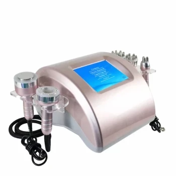
Depth selection should follow the tissue target, not simply the degree of laxity. Superficial fat lies between the dermis and SMAS and is closely associated with skin laxity, while deep fat lies beneath or around facial muscles and contributes more to structural support and gliding. HIFU can place focused thermal injury at deeper planes such as the deep dermis or SMAS, whereas Microneedle RF generally delivers controlled heat within the dermis and superficial subcutaneous tissue. The correct depth depends on regional anatomy, tissue thickness, and the device’s validated treatment parameters.
The key principle is anatomical matching: use superficial settings for dermal remodeling and carefully selected subdermal tightening, and deeper HIFU focal depths when the intended target is the deep dermis or SMAS. Excessive depth or energy in thin areas can reduce valuable facial volume, while insufficient depth may produce little structural improvement.
Why the Two Fat Layers Matter
The superficial fat layer
The superficial fat compartment sits directly beneath the dermis and above the SMAS. Its volume can change with body-weight fluctuations and aging, and its relationship with the overlying skin strongly influences visible laxity.
Heating this region may contribute to subdermal contraction and skin retraction. However, excessive thermal exposure can cause unwanted fat atrophy, particularly in thin facial zones where superficial volume supports contour.
The deep fat layer
Deep facial fat lies beneath or around facial muscles. It contributes to tissue support, smooth gliding between anatomical planes, and overall facial structure.
Deep fat is not simply a larger treatment target. In many facial regions, preserving deep volume is essential because aggressive reduction can worsen hollowing or produce an aged appearance.
Aging changes both layers and their support
With aging, fat may shift between planes, retaining ligaments weaken, and soft tissue descends. This combination contributes to jowls, sagging skin, and deeper facial folds.
Therefore, laxity does not automatically mean that fat should be destroyed. The treatment objective may instead be dermal collagen remodeling, contraction of a fibrous support layer, or preservation of facial volume.
How Anatomy Guides HIFU Depth
HIFU targets a defined focal plane
HIFU creates focused thermal coagulation points at selected depths rather than heating the entire tissue column uniformly. This allows the operator to target the dermis, subcutaneous tissue, or SMAS-region structures while limiting unnecessary superficial injury.
Common cartridge categories include approximately 1.5 mm for dermal remodeling, 3.0 mm for deeper dermal or subcutaneous treatment, and 4.5 mm for the SMAS-region target. These values are device-specific conventions, not universal anatomical guarantees.
Deeper is not automatically better
A 4.5-mm cartridge is appropriate only where the SMAS-region target is actually present at a safe distance from vulnerable structures. In a thin cheek, temple, periorbital area, or other anatomically delicate zone, that depth may be too deep or may place energy near structures that should be preserved.
Similarly, a 3.0-mm setting should not be selected solely because the patient has visible sagging. The operator must establish whether the tissue at that depth is an appropriate target and whether sufficient tissue thickness exists to absorb the energy safely.
HIFU is best suited to structural tightening goals
When the primary objective is lifting or contraction of deeper support tissues, HIFU may be selected because it can create focal thermal points at the deep dermis or SMAS-region plane.
Its role is different from superficial resurfacing. HIFU does not replace treatments designed primarily to improve epidermal texture, pigmentation, or very superficial fine lines.
How Anatomy Guides Microneedle RF Depth
Microneedle RF places energy along the needle path
Microneedle RF delivers radiofrequency energy through insulated or non-insulated needles at a controlled insertion depth. The treatment is therefore usually directed toward the dermis and, depending on the device and setting, the superficial subcutaneous interface.
Depth selection should reflect the thickness of the dermis and superficial fat in the specific region. A setting that is appropriate for a thick lower face may be excessive for the eyelid, temple, or a naturally thin cheek.
RF is primarily a dermal remodeling tool
Microneedle RF is commonly used to create controlled dermal heating that stimulates collagen remodeling, improves elasticity, and may produce some subdermal contraction. It should not be treated as interchangeable with a deep HIFU cartridge aimed at the SMAS.
The practical distinction is important: Microneedle RF generally treats the dermis or superficial subdermal plane, while HIFU can place discrete thermal targets deeper, including the SMAS-region layer when anatomy and the device permit.
Needle depth must match the regional soft-tissue thickness
The operator should avoid selecting needle depth based only on a standardized facial map. Individual variation in skin thickness, fat distribution, age, sex, and prior weight change can substantially alter the distance to deeper structures.
Male skin is often thicker on average, while female skin may be thinner, especially with age-related hormonal changes. These are broad trends rather than substitutes for patient-specific assessment.
The Practical Depth-Matching Framework
Start with the desired biological effect
The first question is what the treatment is intended to change:
- Dermal laxity or fine lines: favor a dermal remodeling strategy.
- Superficial subdermal laxity: consider carefully calibrated superficial or subdermal heating.
- Deeper structural laxity: evaluate whether a deeper HIFU focal plane is appropriate.
- Excess or misplaced volume: recognize that tightening energy may not correct the underlying problem.
A device setting is meaningful only when it corresponds to the intended tissue response.
Map tissue thickness before treating
Clinical examination, including assessment of tissue thickness and a pinch test, can help distinguish superficial from deeper fat. High-resolution ultrasound assessment can provide more direct information about the dermis, fat compartments, muscles, and SMAS-region structures.
This mapping is particularly valuable in thin facial zones, asymmetric faces, previously treated areas, and patients with limited facial volume.
Select the least aggressive effective plane
The safest principle is to use the shallowest or least aggressive setting that can reasonably reach the intended target. The goal is not maximum penetration; it is sufficient energy at the correct anatomical plane.
For example, dermal laxity does not require routine treatment of the SMAS, and superficial skin tightening does not justify unnecessary deep fat heating.
Adjust energy as well as depth
Depth and energy are linked but not identical. A correct depth with excessive energy can still cause thermal injury or unwanted volume loss, while an appropriate energy level delivered too superficially may fail to affect the intended support layer.
Treatment planning must therefore consider cartridge or needle depth, power, pulse duration, density, spacing, and regional anatomy together.
Understanding the Trade-offs
The risk of superficial fat loss
Superficial fat is vulnerable when treatment is too deep, too energetic, too dense, or repeatedly applied. Localized fat atrophy may create hollowing, contour irregularity, or an older appearance rather than a lifted one.
This risk is greatest where the skin and fat compartments are thin or where facial volume is already deficient.
The risk of ineffective treatment
Selecting a depth that is too shallow for the intended structural target may produce only modest skin improvement. Similarly, Microneedle RF cannot be expected to reproduce the effect of a device specifically designed to place HIFU focal points at the SMAS-region plane.
Ineffective treatment often reflects a mismatch between the anatomical problem and the technology, not simply inadequate energy.
Deep anatomical problems may exceed surface-device capabilities
Preplatysmal superficial fat may be accessible to non-surgical tightening approaches in suitable patients. Subplatysmal adiposity, submandibular gland ptosis, or marked platysmal banding are deeper or muscular problems that may not be corrected by surface energy devices.
In these situations, increasing energy or depth is not a reliable solution and may increase risk without addressing the cause.
Standard cartridge labels are not patient anatomy
A nominal 1.5-, 3.0-, or 4.5-mm label describes the device’s intended focal or insertion depth. It does not prove that the patient’s relevant tissue layer lies exactly at that distance in every facial region.
Manufacturers’ instructions, treatment-area restrictions, and validated protocols must take priority over generic depth assumptions.
How to Apply This to Your Project
Treatment planning should connect the patient’s visible concern with the anatomical layer responsible for it.
- If your primary focus is dermal laxity and texture: prioritize a dermal remodeling approach, often using Microneedle RF at a regionally appropriate needle depth or a superficial HIFU setting when indicated.
- If your primary focus is deeper structural lifting: evaluate HIFU focal depths directed toward the deep dermis or SMAS-region layer, using imaging or careful anatomical assessment where available.
- If your primary focus is preserving facial volume: avoid indiscriminate deep or high-energy treatment in thin areas and favor the least aggressive effective settings.
- If your primary focus is localized fat reduction: confirm that volume reduction is actually desirable, because facial fat loss can worsen hollowing and is a different objective from skin tightening.
- If your primary focus is neck or lower-face laxity: distinguish superficial preplatysmal tissue from deeper muscular or subplatysmal problems before selecting a surface energy device.
The best results come from treating the correct anatomical plane with the lowest effective energy while preserving the facial volume and support structures that create a natural contour.
Summary Table:
| Fat Layer | Location | Role | Aging Changes | HIFU Target | RF Target |
|---|---|---|---|---|---|
| Superficial | Between dermis and SMAS | Influences visible laxity and contour | Volume loss or shift, contributes to sagging | Dermal remodeling (1.5–3.0 mm) | Dermal remodeling (needle depth in dermis) |
| Deep | Beneath or around facial muscles | Structural support, gliding | Loss or descent, worsens hollowing | SMAS-region target (4.5 mm) | Not primary target, avoid thermal damage |
Ready to enhance your practice with precise, anatomy-based HIFU and Microneedle RF treatments? BELIS offers professional-grade aesthetic devices for clinics and premium salons, including HIFU and RF platforms. Our experts can guide you on optimal depth selection and treatment protocols. Contact us today to learn more.
Related Products
- 4D 12D HIFU Machine Device for Skin Tightening
- 4D 12D HIFU Machine Device for Skin Tightening and Lifting
- 22D HIFU Machine Device Facial Machine
- 7D 12D 4D HIFU Machine Device
- 12D HIFU Machine Device for Facial HIFU Treatment
People Also Ask
- How do non-invasive skin tightening devices such as HIFU and microneedle RF compare to surgical rhytidectomy for facial lifting? Discover the key differences in results, longevity, and recovery.
- How does intense ultrasound (HIFU) skin tightening act on facial soft tissues? Discover its key clinical contraindications.
- What is the mechanism of action of High-Intensity Focused Ultrasound (HIFU) devices in noninvasive body sculpting, and how is surrounding tissue protected?
- How do non-invasive lifting devices like high-intensity focused ultrasound (HIFU) complement soft tissue fillers in treating lower face aging and pre-jowl sulcus depression? Discover the synergistic approach.
- What key clinical safety guidelines and contraindications must aesthetic practitioners observe when operating ultrasound skin tightening devices? Essential Safety Protocols for Clinics