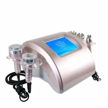
The integration of dermoscopy into High-Intensity Focused Ultrasound (HIFU) systems provides real-time optical surveillance that transforms a traditionally "blind" procedure into a precision-guided intervention. This technology allows clinicians to identify microscopic biomarkers of sebaceous hyperplasia, track immediate tissue responses to acoustic energy, and ensure that treatment is localized to the lesion while sparing the surrounding healthy dermis.
Core Takeaway: Integrated dermoscopy bridges the gap between diagnostic visualization and therapeutic delivery, enabling clinicians to monitor "crown vessels" and immediate tissue whitening to maximize surgical precision and patient safety during HIFU procedures.
Enhancing Diagnostic Precision and Parameter Selection
Identifying Pathognomonic Markers
Sebaceous hyperplasia presents with specific dermatoscopic structures, such as crown vessels and cloud-like signs, which are difficult to distinguish with the naked eye. The integrated system allows the clinician to confirm the diagnosis at a microscopic level immediately before the ultrasound pulse is delivered.
Customizing Treatment Dosimetry
High-precision digital images serve as the foundational data for determining personalized HIFU parameters. By assessing the size and vascularity of the lesion via the dermatoscope, the operator can more accurately calibrate the power and pulse duration required for effective clearance.
Establishing a Clinical Baseline
Capturing high-resolution images prior to treatment provides a vital reference point for the healing process. These baseline images allow for objective comparison during post-treatment follow-ups to verify complete lesion resolution.
Real-Time Intraoperative Monitoring
Visualizing Thermal and Mechanical Responses
As HIFU delivers energy, the integrated camera allows the clinician to observe immediate physical reactions on a user interface. Signs such as local whitening, swelling, or shrinkage indicate that the energy has reached the therapeutic threshold, allowing for "on-the-fly" adjustments.
Tracking Acoustic Cavitation
HIFU relies on acoustic cavitation, where microbubbles oscillate and collapse to disrupt cellular structures through mechanical shear forces. Real-time monitoring ensures these mechanical effects are occurring within the intended target zone, complementing the thermal ablation process for more thorough clearance.
Ensuring Alignment via Red-Dot Guidance
The integrated system often utilizes a red-dot guide on the digital screen to align the ultrasound transducer with the target. This visual feedback mechanism is critical for ensuring that every pulse effectively acts on the sebaceous gland without damaging adjacent healthy tissue.
Understanding the Trade-offs and Limitations
Cognitive Load and Procedural Speed
While real-time monitoring increases precision, it also increases the cognitive load on the operator, who must simultaneously monitor the screen and the patient. This can lead to slightly longer procedure times compared to traditional, non-monitored thermal treatments.
Limitations of Surface Visualization
Dermoscopy provides a high-resolution view of the skin's surface and upper dermis, but it cannot fully visualize the depth of ultrasound penetration. Operators must still rely on their technical knowledge of the device's focal depth, as the visual feedback is a proxy for, rather than a direct view of, deep subcutaneous changes.
Equipment Sensitivity and Maintenance
Integrated optical systems add a layer of complexity to the HIFU handpiece. These sensitive digital components require rigorous maintenance and calibration to ensure that the visual alignment remains perfectly synchronized with the ultrasound focal point.
How to Apply This to Your Practice
The use of integrated dermoscopy is essential for clinicians who prioritize precision and want to minimize the risk of scarring or incomplete treatment.
- If your primary focus is Clinical Safety: Use the real-time visual feedback to stop energy delivery the moment "tissue whitening" is observed, preventing over-treatment.
- If your primary focus is Aesthetic Outcomes: Rely on the identification of "crown vessels" to ensure the ultrasound focal point is centered exactly on the hyperplasia, reducing collateral damage to healthy skin.
- If your primary focus is Patient Documentation: Utilize the integrated camera to capture standardized pre- and post-operative images for medical records and treatment validation.
The integration of real-time optical monitoring represents the next evolution in non-invasive surgery, providing the visual clarity necessary to harness the full potential of high-intensity ultrasound.
Summary Table:
| Feature | Benefit for Clinicians | Clinical Impact |
|---|---|---|
| Real-time Optical Monitoring | Visualizes "crown vessels" & tissue response | Prevents "blind" procedures; ensures accuracy |
| Red-Dot Guidance | Precise alignment of ultrasound transducer | Minimizes damage to surrounding healthy tissue |
| Personalized Dosimetry | Customizes power/pulse based on lesion size | Higher clearance rates with fewer side effects |
| Digital Documentation | Automated pre/post-treatment image capture | Enhances follow-up accuracy and patient trust |
Elevate Your Clinic’s Precision with BELIS Medical Aesthetics
At BELIS, we specialize in providing professional-grade medical aesthetic equipment designed exclusively for clinics and premium salons. Whether you are treating complex skin conditions like Sebaceous Hyperplasia or offering body contouring, our advanced systems ensure safety and superior results.
Our Professional Portfolio Includes:
- Advanced Laser Systems: Diode Hair Removal, Alexandrite, CO2 Fractional, Erbium, Nd:YAG, and Pico lasers.
- Precision Lifting & Tightening: State-of-the-art HIFU and Microneedle RF technology.
- Body Sculpting Solutions: EMSlim, Cryolipolysis, and RF Cavitation.
- Specialized Care: Hydrafacial systems, high-tech skin testers, and hair growth machines.
Ready to integrate high-precision technology into your practice? Contact our specialists today to discuss how BELIS can enhance your clinical outcomes and business growth.
References
- Bartosz Woźniak, Jacek Calik. Treatment of Sebaceous Hyperplasia by High-Frequency Focused Ultrasound (HIFU): A Comprehensive Exploration with Clinical Insights. DOI: 10.3390/jcm14041305
This article is also based on technical information from Belislaser Knowledge Base .
Related Products
- 22D HIFU Machine Device Facial Machine
- 4D 12D HIFU Machine Device for Skin Tightening and Lifting
- 4D 12D HIFU Machine Device for Skin Tightening
- 12D HIFU Machine Device for Facial HIFU Treatment
- 7D 12D 4D HIFU Machine Device
People Also Ask
- Is swelling a frequent side effect after a HIFU facial? Expert Insights on HIFU Recovery and Safety
- What is the mechanism of action of High-Intensity Focused Ultrasound (HIFU) devices in noninvasive body sculpting, and how is surrounding tissue protected?
- How do High-Intensity Focused Ultrasound (HIFU) devices function? Master Non-Invasive Face Lifting Technology
- Are home-use HIFU devices a recommended alternative? Why Clinical Safety Beats At-Home Risks
- What are the main functions of a High Intensity Focused Ultrasound (HIFU) machine? Advanced Skin Lifting and Sculpting