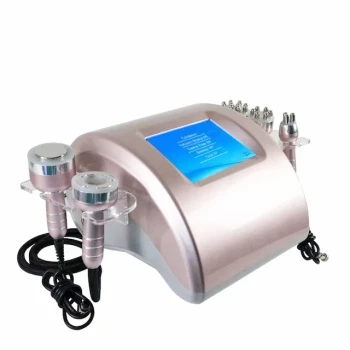
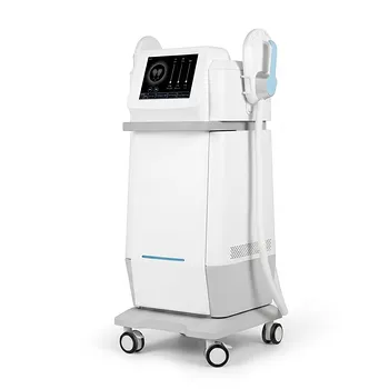
Clinics should treat the actinic keratosis first, then sequence resurfacing and tightening according to lesion status, skin condition, and laxity. A practical plan separates medical management of sun-damaged or potentially precancerous lesions from cosmetic correction of sagging. After appropriate evaluation, fractional laser resurfacing may address selected superficial photodamage, while HIFU, microneedle RF, or deep infrared systems target dermal laxity and collagen remodeling.
The safest structure is staged, not simultaneous by default: diagnose and manage actinic keratoses first, restore the skin barrier, then introduce resurfacing and deeper tightening with conservative parameters and documented clinical endpoints.
Start With Medical Risk Stratification
Confirm that the lesion is appropriate for cosmetic treatment
Actinic keratosis is a premalignant keratinocytic lesion, not simply an aesthetic rough spot. A clinician should assess whether each lesion is typical, recurrent, thick, tender, bleeding, rapidly changing, or otherwise suspicious for progression to squamous cell carcinoma.
Suspicious or uncertain lesions should be referred for dermatologic assessment and biopsy when indicated. Cosmetic laser or tightening treatment should not be used to obscure, delay, or empirically treat a lesion that requires diagnostic evaluation.
Map the treatment areas
Document the distribution and severity of:
- Actinic keratoses and diffuse field damage
- Erythema, pigmentation, and telangiectasia
- Fine and deep wrinkles
- Skin thickness and dryness
- Laxity of the cheeks, jawline, submental region, and neck
- Previous procedures, scarring, and pigmentary response
This mapping helps the clinic distinguish surface disease, photodamage, and structural laxity rather than treating the entire face with one device.
Establish patient-specific safety factors
The consultation should review skin phototype, tanning or recent sun exposure, wound-healing history, herpes simplex history when relevant, medications, autoimmune disease, active dermatitis, and prior energy-based treatments.
The patient should also understand that tightening devices generally produce gradual improvement, while ablative resurfacing involves more downtime and a higher risk of erythema, pigment alteration, infection, and prolonged healing.
Build the Plan Around Treatment Depth
Treat actinic keratosis and field cancerization first
The initial phase should focus on medical management of visible lesions and broader sun-damaged fields. Depending on the clinical assessment, this may include established dermatologic treatments such as cryotherapy, prescription field therapy, photodynamic therapy, or another physician-selected approach.
Fractional ablative CO₂ or Er:YAG resurfacing can be considered for selected superficial photodamage and texture concerns, but it should not replace appropriate diagnosis or standard management of suspicious actinic keratoses.
Restore the epidermal barrier before aggressive resurfacing
Patients aged 47 to 56 commonly have drier skin and reduced tolerance for aggressive treatment. Clinics should first control inflammation, optimize moisturization, reinforce photoprotection, and allow treated areas to heal fully.
A stable barrier improves comfort and reduces the likelihood that subsequent resurfacing or tightening will be performed on compromised skin.
Address surface texture with fractional laser systems
Fractional ablative lasers create controlled columns of thermal injury to improve epidermal irregularity, fine lines, photodamage, and selected scars. CO₂ systems generally provide more aggressive resurfacing, while Er:YAG systems can offer a more superficial ablation profile.
The choice should be based on lesion status, wrinkle depth, skin type, downtime tolerance, and the operator’s ability to manage complications. Treatment should be conservative around recently treated lesions and postponed if healing is incomplete.
Address laxity with deeper energy-based devices
HIFU, microneedle RF, and deep infrared fractional systems are intended to deliver thermal energy below the epidermis, promoting collagen contraction and longer-term remodeling. They are most relevant to laxity along the jawline, lower face, submental area, and neck.
These devices improve tissue firmness but do not remove substantial excess skin. Patients with advanced structural laxity should be counseled that surgery may provide a more predictable lift, while energy-based treatment may serve as a non-surgical or maintenance option.
Sequence the Modalities Rather Than Stacking Them
Use a staged treatment pathway
A reasonable clinical sequence is:
- Assessment and lesion management
- Barrier repair and sun-protection preparation
- Targeted resurfacing for texture and photodamage
- Deeper tightening after the skin has recovered
- Maintenance and surveillance
The exact interval should be determined by healing, inflammation, device parameters, and the patient’s response. A fixed calendar should not override clinical recovery.
Consider a multi-layer protocol
For suitable patients, clinics may combine complementary modalities over separate sessions:
- Fractional ablative laser: epidermal and upper-dermal resurfacing
- Deep infrared fractional energy: deeper dermal remodeling and laxity treatment
- HIFU or RF: focused tightening of selected facial and neck zones
- IPL: dyschromia and vascular concerns when appropriately diagnosed
IPL may improve redness and pigmentation, but it should not be presented as a primary treatment for actinic keratosis or as a substitute for lesion-directed medical care.
Use conservative treatment zones
Avoid treating every area with maximum energy. Thin periocular skin, recently inflamed skin, active dermatitis, and incompletely healed areas require particular caution.
A zone-based plan is usually more defensible: treat medical lesions according to their clinical needs, resurface photodamaged areas selectively, and tighten only where laxity is the principal concern.
Set Realistic Expectations for Results
Explain the different timelines
Resurfacing can improve texture and fine lines as healing progresses, whereas tightening is usually more gradual. Patients may notice some early contraction, but the visible result can continue developing over several months as collagen remodeling proceeds.
A common multi-session framework is three to five sessions spaced approximately one month apart for selected fractional or light-based protocols, but this should be adapted to the modality, intensity, and recovery pattern.
Define success by separate endpoints
The clinic should assess:
- Reduction in clinically identified actinic keratoses
- Improvement in roughness and photodamage
- Smoother wrinkles and surface texture
- Improved jawline or neck definition
- Patient-reported firmness and satisfaction
- Absence of prolonged adverse effects
Separating these endpoints prevents a patient from judging a medical lesion treatment solely by cosmetic lifting, or judging a tightening procedure by surface smoothness alone.
Include surveillance in the plan
Actinic keratoses indicate cumulative ultraviolet damage and ongoing risk. Continued dermatologic surveillance, daily broad-spectrum sunscreen use, protective clothing, and avoidance of tanning are essential even after successful cosmetic treatment.
New, persistent, thickening, bleeding, or painful lesions should be reassessed rather than repeatedly treated as cosmetic irregularities.
Understanding the Trade-offs
Ablative resurfacing offers stronger surface correction
Ablative fractional laser can provide substantial improvement in photodamage and texture, but it creates a controlled wound. The trade-offs include downtime, discomfort, infection risk, post-inflammatory pigment alteration, and the need for disciplined aftercare.
The risk profile is particularly important in patients with darker skin phototypes, a history of poor wound healing, or unrealistic expectations about recovery.
Tightening devices have limits
HIFU, RF, and infrared systems may improve mild to moderate laxity, but their results are not equivalent to excisional surgery. Excess skin, pronounced jowling, or severe neck laxity may not respond adequately to non-surgical tightening alone.
Microneedle RF also combines mechanical penetration with thermal injury, so treatment should be avoided or modified when the skin is actively inflamed, infected, or not fully healed.
More devices do not automatically mean better outcomes
Combining multiple energy sources can increase cumulative inflammation without producing proportional benefit. Clinics should have a clear reason for each modality, use staged recovery periods, and avoid treating the same area aggressively simply because several devices are available.
Do not compromise lesion diagnosis for cosmetic goals
The most important limitation is clinical prioritization. A smoother or tighter appearance is not a successful outcome if a suspicious keratinocytic lesion was missed, inadequately evaluated, or masked by cosmetic treatment.
How to Apply This to Your Project
The final protocol should be individualized by a qualified clinician and documented as a medical treatment pathway, not merely as a device package.
- If your primary focus is actinic keratosis safety: Complete lesion assessment and appropriate medical treatment first, and biopsy or refer any suspicious lesion before cosmetic energy procedures.
- If your primary focus is surface photodamage: Use selected fractional CO₂ or Er:YAG resurfacing only after the skin is stable, with parameters matched to phototype, lesion status, and recovery capacity.
- If your primary focus is facial or neck laxity: Use HIFU, microneedle RF, or deep infrared treatment for appropriately selected mild-to-moderate laxity, while explaining that remodeling is gradual and excess skin may require surgery.
- If your primary focus is comprehensive rejuvenation: Sequence modalities by depth—medical lesion care, epidermal resurfacing, then deeper tightening—rather than performing aggressive treatments concurrently by default.
- If your primary focus is long-term outcomes: Build in sun protection, maintenance treatments, outcome photography, and ongoing dermatologic surveillance for new or recurrent lesions.
A well-structured plan treats the skin’s medical risk first, then addresses texture and laxity in separate, measurable stages.
Summary Table:
| Stage | Focus | Treatment Options | Goal |
|---|---|---|---|
| 1 | Medical Risk Management | Cryotherapy, PDT, field therapy | Manage actinic keratosis and field cancerization safely |
| 2 | Barrier Repair & Prep | Moisturizers, sun protection | Restore skin barrier before cosmetic procedures |
| 3 | Surface Resurfacing | Fractional CO2 or Er:YAG laser | Improve texture, photodamage, fine lines |
| 4 | Deep Tightening | HIFU, Microneedle RF, Deep Infrared | Address laxity and collagen remodeling |
| 5 | Maintenance & Surveillance | Sunscreen, follow-ups | Maintain results and monitor for new lesions |
Elevate your clinic's aesthetic offerings with BELIS's professional-grade devices, including advanced lasers, IPL, PDT, HIFU, and body contouring systems. Our solutions are designed exclusively for clinics and premium salons, ensuring safety, efficacy, and patient satisfaction. For distributors, we offer robust OEM/ODM support, certified quality, and reliable supply chains to boost your business. Contact us today to learn how BELIS can help you deliver comprehensive, staged treatments that prioritize patient safety and outstanding results.
Related Products
- 4D 12D HIFU Machine Device for Skin Tightening
- 4D 12D HIFU Machine Device for Skin Tightening and Lifting
- 22D HIFU Machine Device Facial Machine
- Ultrasonic Cavitation Radiofrecuency Machine for Body Slimming
- 7D 12D 4D HIFU Machine Device
People Also Ask
- How do non-invasive skin tightening devices such as HIFU and microneedle RF compare to surgical rhytidectomy for facial lifting? Discover the key differences in results, longevity, and recovery.
- What is the mechanism of action of High-Intensity Focused Ultrasound (HIFU) devices in noninvasive body sculpting, and how is surrounding tissue protected?
- How do non-invasive lifting devices like high-intensity focused ultrasound (HIFU) complement soft tissue fillers in treating lower face aging and pre-jowl sulcus depression? Discover the synergistic approach.
- What key clinical safety guidelines and contraindications must aesthetic practitioners observe when operating ultrasound skin tightening devices? Essential Safety Protocols for Clinics
- Why is it necessary to fill the transducer chamber with deionized water and PE film in HIFU devices? Key Safety Insights