For Port Wine Stains (PWS), treatment should be individualized rather than guided by a single universal setting. Clinicians commonly begin with a test patch, use conservative fluence, and assess the response after 6–8 weeks before modifying parameters. For a typical 7 mm spot size, initial fluences are often approximately 4.5–8 J/cm², with lower starting values for children and sensitive anatomical areas.
The safest protocol is incremental: test first, reassess after adequate healing, and increase fluence only when the tissue response supports it. Pain control should combine topical anesthesia, cooling, and—when necessary—age-appropriate sedation or general anesthesia with complete ocular protection.
Establishing the Treatment Parameters
Begin with a test patch
A fluence test patch helps determine how the patient’s skin and lesion respond before treating the entire PWS. Clearing and adverse effects should generally be evaluated 6–8 weeks after treatment, rather than immediately.
The treatment response, including lesion lightening and any blistering, prolonged erythema, pigment alteration, or scarring, should guide subsequent settings.
Select a conservative starting fluence
With a commonly used 7 mm spot size, initial fluence may fall in the range of 4.5–8 J/cm². The lower end is generally more appropriate for pediatric patients, sensitive areas, darker skin types, or other situations with increased epidermal injury risk.
For pulsed-dye laser treatment near 595 nm, some protocols use approximately 6.5–8.5 J/cm², spot sizes of 10–12 mm, and pulse durations near 0.5 milliseconds. These figures are device- and patient-dependent and should not replace manufacturer guidance or clinical judgment.
Increase energy gradually
If the lesion responds without unacceptable tissue injury, fluence may be increased in small increments, commonly 0.25–0.5 J/cm². Pediatric protocols described in the references often begin around 5.0–5.5 J/cm² and increase by approximately 0.5 J/cm² per session.
A non-responding area should not simply be treated repeatedly at higher energy. Persistent nonresponse may indicate deeper or larger vessels, inadequate wavelength selection, or an unfavorable lesion location rather than a need for aggressive escalation.
Match the Laser to Lesion Depth
Superficial pink or red lesions
Flat, superficial PWS lesions generally respond best to vascular dye lasers that preferentially target hemoglobin while limiting melanin absorption. Short-pulse treatment and appropriate cooling can produce vascular injury while reducing epidermal damage.
Treatment is usually performed over multiple sessions, often spaced 3–4 months apart, allowing tissue recovery and gradual vascular clearance.
Thick, dark, or nodular lesions
Thicker, darker, or nodular lesions may contain larger or deeper vessels that are less responsive to conventional single-wavelength treatment. Approximately 20–30% of PWS may be resistant to these treatments for this reason.
For recalcitrant lesions, clinicians may consider longer-wavelength systems such as 755 nm Alexandrite, 810 nm diode, or 1064 nm Nd:YAG lasers. These wavelengths penetrate more deeply, but they also require particularly careful fluence, pulse-duration, and cooling control because of the greater risk of nonspecific thermal injury.
Consider anatomy and lesion characteristics
Treatment planning should account for:
- Anatomical location: Face, neck, chest, upper arms, lateral cheek, and forehead lesions often respond better than lesions on the hands or legs.
- Lesion size: Smaller lesions, particularly those under approximately 20 cm², often clear more readily.
- Vessel depth and thickness: Superficial lesions are more suitable for dye-laser treatment, whereas deeper or hypertrophic lesions may require a longer-wavelength approach.
These factors influence the choice of wavelength, spot size, pulse duration, fluence, and expected number of sessions.
Use Cooling and Controlled Delivery
Protect the epidermis with cooling
External or dynamic cooling should accompany vascular laser treatment when appropriate. Cooling protects the epidermis, reduces treatment discomfort, and may decrease post-treatment purpura, hyperpigmentation, and downtime.
For longer-wavelength treatment, cooling is especially important because deeper penetration can increase the risk of epidermal and dermal thermal injury.
Avoid excessive heat accumulation
Treatment delivery should prevent overlapping energy from creating excessive local heating. Computerized scanning systems may use non-adjacent treatment spots or controlled grids to distribute energy more uniformly and reduce geometric patterning.
Double-pass techniques may be considered in selected cases, but only with carefully adjusted fluence and timing. Uncontrolled overlap increases the risk of blistering, scarring, and post-inflammatory hyperpigmentation.
Treat resistant regions strategically
When superficial treatment produces incomplete clearing, changing the treatment strategy is preferable to repeatedly increasing energy over the same nonresponsive area. A deeper-penetrating laser or a carefully selected combination approach may be more rational after reassessing vessel depth and lesion morphology.
Pain Management Protocols
Use topical anesthetic when appropriate
For cooperative patients, topical anesthetic creams can reduce discomfort:
- EMLA: 2.5% lidocaine and 2.5% prilocaine, commonly applied under occlusion for approximately 90 minutes to 4 hours.
- Amethocaine 4% gel: commonly applied for approximately 30–45 minutes.
Application time, occlusion, treated surface area, patient age, and medical history must be considered. These agents should be used according to local prescribing requirements and product instructions.
Add cooling during laser delivery
Cooling is both a pain-control measure and a safety measure. It can reduce the heat sensation during pulses and limit epidermal injury.
Cooling should be coordinated with the laser’s wavelength and treatment window rather than used as a substitute for appropriate fluence selection.
Plan sedation for children when necessary
Young children may not tolerate the duration, noise, discomfort, or need to remain still during treatment. Pediatric cases may therefore require procedural sedation or general anesthesia, depending on lesion size, location, age, treatment setting, and institutional policy.
If general anesthesia or sedation is used, the procedure requires appropriate monitoring and a qualified anesthesia team. Anesthesia does not eliminate the need for conservative laser parameters or careful postoperative observation.
Protect the eyes completely
Ocular protection is mandatory for treatments near the eyes and should be used whenever laser pulses could expose ocular structures. Pediatric patients receiving sedation or general anesthesia require particular attention because they cannot reliably respond to discomfort or reposition themselves.
Protection must be appropriate for the laser wavelength and applied according to established laser-safety protocols.
Assess Treatment Response Before Repeating
Allow adequate recovery
Sessions are commonly spaced 3–4 months apart, although the interval should reflect the device, treatment reaction, lesion location, and degree of tissue recovery. Treating too frequently can compound inflammation and pigmentary or textural complications.
Define a favorable response
Expected response is gradual lightening of the PWS over successive treatments. The clinician should document standardized photographs, lesion color, thickness, treatment settings, and adverse effects to distinguish true nonresponse from insufficient healing time.
Recognize warning signs
Persistent blistering, ulceration, severe pain, prolonged crusting, scarring, or significant pigmentary change indicates that the protocol requires reassessment. Further treatment should generally be deferred until the skin has healed and the cause of the reaction has been evaluated.
Understanding the Trade-offs
Higher fluence is not automatically better
Increasing fluence may improve vascular coagulation, but it also raises the risk of blistering, scarring, dyspigmentation, and nonspecific coagulative necrosis. Incremental adjustment is safer than aggressive escalation.
Deeper penetration increases both capability and risk
Longer wavelengths can reach deeper or larger vessels, making them useful for thick or resistant PWS. However, the same deeper energy delivery can cause more substantial thermal injury if pulse parameters, cooling, or skin selection are inappropriate.
Early treatment may improve practicality
Infant lesions may have smaller, more superficial vessels and can sometimes achieve clearance in fewer sessions when treatment begins early. Nevertheless, early treatment requires careful consideration of anesthesia, ocular protection, skin maturity, and the risks and benefits for the individual child.
Protocols are device-specific
Fluence, spot size, pulse duration, cooling method, and endpoint cannot be transferred directly from one laser platform to another. The figures above are clinical reference ranges, not universal prescriptions.
How to Apply This to Your Project
The following framework can help organize a clinician-led treatment plan:
- If your primary focus is initial safety: Perform a test patch, begin conservatively, use cooling, and reassess the response at 6–8 weeks before changing fluence.
- If your primary focus is pediatric treatment: Consider lower starting fluence—often around 5.0–5.5 J/cm² with a 7 mm spot—and plan topical anesthesia, sedation, or general anesthesia according to the child’s needs.
- If your primary focus is pain reduction: Combine topical anesthetic with active cooling, and use monitored sedation or general anesthesia when immobility or procedural tolerance requires it.
- If your primary focus is a thick or resistant PWS: Reassess vessel depth and consider a longer-wavelength option such as Alexandrite or Nd:YAG rather than repeatedly overtreating the same area.
- If your primary focus is preventing complications: Use wavelength-specific ocular protection, avoid excessive overlap, document treatment responses, and stop escalation when tissue injury appears.
A safe PWS protocol is built on measured energy delivery, adequate recovery time, effective analgesia, and regular reassessment—not on maximizing fluence.
Summary Table:
| Parameter/Protocol | Recommendation |
|---|---|
| Initial Fluence (7 mm spot) | 4.5–8 J/cm², lower for children/sensitive areas |
| Test Patch | Perform before full treatment; reassess at 6–8 weeks |
| Fluence Increments | 0.25–0.5 J/cm² per session if tolerated |
| Treatment Interval | 3–4 months between sessions |
| Cooling | Use active or dynamic cooling to protect epidermis and reduce pain |
| Topical Anesthetics | EMLA (90 min–4 h) or Amethocaine 4% gel (30–45 min) |
| Pediatric Sedation | Consider sedation or general anesthesia for young children |
| Ocular Protection | Mandatory for all treatments, especially near eyes |
| Resistant Lesions | Consider longer wavelengths (755 nm, 810 nm, 1064 nm) |
Ready to Enhance Your PWS Laser Treatments?
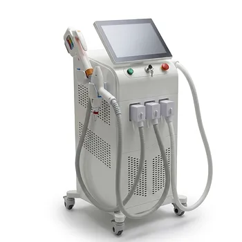
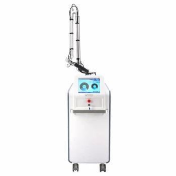
At BELIS, we provide advanced vascular laser systems (including pulsed dye, Alexandrite, and Nd:YAG) designed for safe and effective treatment of Port Wine Stains. Our equipment features precise energy control, integrated cooling, and pediatric-friendly options to help you achieve optimal outcomes while prioritizing patient comfort.
Partner with us to access:
- State-of-the-art laser technology tailored for clinical excellence
- Comprehensive training and after-sales support
- Customizable solutions to meet the unique needs of your practice
Contact us today to learn how BELIS can elevate your PWS treatments and grow your aesthetic practice.
Related Products
- 808nm Diode Laser Hair Removal Machine 755+808+1064nm Mixed Wavelength Professional Equipment
- Clinic Use IPL and SHR Hair Removal Machine with Nd Yag Laser Tattoo Removal
- Clinic Use IPL SHR ND YAG Laser Hair Removal RF Skin Tightening Machine
- Hydrofacial Machine with Facial Skin Analyzer and Skin Tester
- Pico Laser Tattoo Removal Machine Picosure Picosecond Laser Machine
People Also Ask
- How do 800/810 nm diode hair removal lasers compare to 1064 nm long-pulsed Nd:YAG lasers? Discover the Best for Your Patients
- What ocular safety protocols are required when operating diode or alexandrite hair removal lasers near the periorbital area? Essential eye protection and shielding for safe treatment.
- How can aesthetic practitioners prevent side effects like paradoxical hair regrowth and thermal burns when performing diode laser hair removal on dark skin? Master safe protocols for Fitzpatrick IV–VI skin.
- What is the recommended treatment interval for diode laser hair removal? Achieve Max Hair Reduction with 45-Day Sessions
- How do broad-spectrum noncoherent light sources compare to single-wavelength diode lasers in aesthetic hair removal applications? Find the best fit for your clinic.