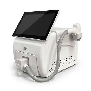
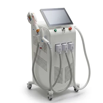
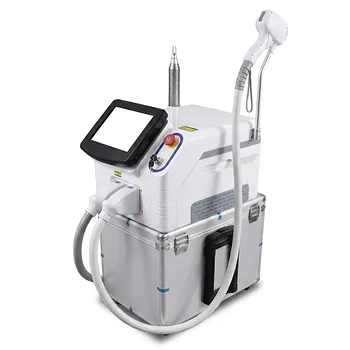
Thermal safety depends on controlling cumulative heat, not merely selecting a single safe pulse energy. Long-pulse 1064 nm Nd:YAG lasers reach deep dermal vessels and hair follicles while sparing relatively melanin-rich epidermis, but their low absorption in water produces broad, slowly cooling thermal volumes. The principal risks are heat accumulation, epidermal injury, blistering, scarring, pigmentary change, dermal necrosis, and—at depth—nerve injury.
Core takeaway: Use active epidermal cooling, avoid pulse stacking, and allow adequate inter-pulse cooling. Because 1064 nm energy penetrates deeply and can produce delayed cumulative heating, treatment endpoints and tissue response must be monitored rather than inferred from a single pulse parameter.
Why 1064 nm requires deliberate thermal management
Deep penetration creates a large thermal volume
Long-pulse 1064 nm light can penetrate approximately several millimeters into the dermis. This supports treatment of deep veins and deeply rooted hair follicles, but it also exposes a relatively large volume of normal tissue to heat.
The wavelength has comparatively low absorption by epidermal melanin and water. Energy therefore spreads more broadly and cools more slowly than it would in a strongly absorbing, superficial target.
High fluence increases the margin for error
Hemoglobin absorption is relatively low at 1064 nm, so vascular treatment may require higher fluences than shorter vascular wavelengths. The additional energy is also absorbed by surrounding tissue water and can increase the risk of nonspecific heating.
Fluence, pulse duration, spot size, repetition rate, and tissue thickness must therefore be considered together. A parameter that is appropriate for one vessel caliber or hair density should not automatically be transferred to another indication.
Thermal damage is nonlinear
Protein denaturation accelerates rapidly once tissue reaches critical temperature ranges, approximately 60°C to 85°C according to the referenced damage model. This means tissue can appear clinically acceptable during early pulses and then undergo a rapid transition to full-thickness thermal injury after additional heat accumulates.
A small increase in cumulative temperature may therefore produce a disproportionately large increase in damage.
Cooling and pulse timing are the primary safeguards
Use active epidermal cooling
Surface contact cooling, dynamic cryogen cooling, or integrated cold-air cooling should be used when delivering high-fluence long-pulse treatments. Cooling protects the epidermis while allowing the deeper target to remain heated.
Cooling must be synchronized with laser delivery and applied consistently across the treatment area. It should not be used to justify excessive fluence, prolonged exposure, or inadequate spacing between pulses.
Avoid pulse stacking
Pulse stacking is a major thermal hazard, particularly in vascular treatment where high fluences may be required. Repeated pulses delivered to the same location before heat has dissipated can cause bulk temperature accumulation and sudden epidermal destruction.
Possible consequences include:
- Vesiculation and blistering
- Epidermal sloughing or ulceration
- Eschar formation
- Dermal necrosis and scarring
- Tissue atrophy
- Prolonged postoperative pain
A clear treatment grid, deliberate handpiece movement, and documented pulse spacing help prevent accidental repeated exposure.
Set inter-pulse intervals according to cooling
There is no universally safe inter-pulse interval for every device, skin type, target, or anatomical site. The interval should be long enough to prevent clinically meaningful heat accumulation, especially when using large spots, high fluence, or dense pulse delivery.
High repetition rates should be approached cautiously because the treated area may retain heat across multiple pulses. Lower repetition rates and longer rest intervals are particularly important when treating thick tissue, poorly perfused areas, or regions where cooling is less effective.
Match thermal exposure to the treatment target
Vascular coagulation
For vessels, pulse duration should be related to vessel diameter and thermal relaxation behavior. The supplementary reference gives examples of approximately 25 ms for 1 mm vessels and 50 ms for 2–3 mm reticular veins, but these should be treated as protocol examples rather than universal prescriptions.
Larger or deeper vessels generally require different pulse timing and energy delivery than superficial telangiectasia. Spot size should likewise reflect vessel depth and caliber; reported examples range from smaller spots for telangiectasia to larger spots for reticular veins.
The desired endpoint is controlled vascular coagulation, not surface whitening, blistering, or excessive epidermal reaction. A visible endpoint should be interpreted together with patient discomfort, cooling performance, skin type, and the possibility of delayed deep injury.
Hair removal
For hair removal, the target is the deeply located follicular structure rather than the epidermis. Long-pulse 1064 nm treatment is useful because it reaches deeper follicles and has lower melanin absorption than shorter wavelengths, which can offer a broader safety margin in darker skin types.
That margin is not absolute. Excessive fluence, overlapping pulses, insufficient cooling, or treating recently tanned or highly pigmented skin can still cause epidermal heating and injury.
Deep or thick tissue
Dense tissue may require greater energy density or longer exposure to reach a therapeutic thermal threshold. However, the supplementary reference’s example of 35–40 J/cm², approximately 35 ms, and 1 Hz is indication- and device-specific and should not be generalized to all vascular or hair-removal procedures.
The correct approach is to calibrate treatment to tissue thickness, target depth, perfusion, skin type, and the manufacturer’s validated protocol. Higher energy should be accompanied by more conservative pulse spacing and reliable active cooling—not simply faster delivery.
Monitor the treatment zone, not only the laser settings
Assess immediate clinical endpoints
Operators should inspect the treated area continuously for excessive erythema, epidermal whitening, blistering, char, tissue sloughing, or sharply increasing pain. These findings indicate that thermal exposure may be exceeding the intended therapeutic range.
Pain is an important warning signal, but it is not a complete safety monitor. Deep heating may be significant before surface findings become obvious.
Account for delayed thermal injury
Because the 1064 nm wavelength heats a relatively large volume that cools slowly, injury may evolve after treatment. Follow-up assessment should consider delayed blistering, ulceration, persistent pain, pigmentary change, induration, or scarring.
When treating deep or complex lesions, real-time methods such as ultrasound may help define the thermal coagulation zone where clinically available and appropriate. Monitoring is especially valuable when surface appearance does not reliably indicate the depth of heating.
Control exposure duration
Thermal injury is governed by output power, exposure duration, and rest intervals. The supplementary reference advises limiting continuous exposure at average energy levels of approximately 20–50 W to under five seconds, with greater caution above 50 W unless active cooling and longer rest periods are used.
These figures are not a substitute for device-specific validation. They illustrate the principle that average power and cumulative dwell time matter as much as nominal pulse energy.
Protect high-risk anatomical structures
Avoid neural structures
Deep penetration makes nerve injury possible when energy is delivered near major motor nerves or confined anatomical spaces. High-risk areas, including the parotid region containing the facial nerve, require particular caution or avoidance.
Treatment should be conservative in anatomically crowded areas, and the operator should not assume that intact skin means deeper structures are protected.
Use special care for mucosal or interstitial delivery
Transmucosal and interstitial techniques require different thermal controls from noninvasive skin treatment. The reference describes use of bare quartz fibers in non-contact or minimally invasive modes, with low-power settings and short exposures.
The fiber tip should remain in continuous motion across multiple passes and directions rather than dwelling at one location. This reduces the formation of unpredictable focal hot spots.
Respect orbital boundaries
Facial vascular treatment should avoid areas inside the orbital rim. Appropriate ocular protection and strict adherence to laser safety procedures remain essential, because eye injury is a separate and potentially catastrophic hazard from tissue heating.
Understanding the Trade-offs
More energy is not automatically better
Higher fluence may be needed at 1064 nm because of lower hemoglobin absorption, but it also increases nonspecific heating. Raising energy without improving cooling or pulse spacing can convert a controlled treatment into a burn.
Cooling protects the surface but not necessarily deep tissue
Surface cooling can preserve epidermal integrity while deeper tissue continues to accumulate heat. Operators must therefore manage both surface temperature and deep cumulative exposure.
A cool skin surface does not prove that the dermis or follicle is within a safe thermal range.
Longer pulses and faster repetition have different risks
A longer pulse can distribute energy over a greater period, while rapid repetition can build heat before the tissue cools. Neither parameter is inherently safe or unsafe in isolation.
Pulse duration, fluence, repetition rate, spot size, tissue perfusion, and cooling must be evaluated as a combined thermal system.
Do not rely on generic protocol numbers
Published or supplied parameter examples vary by device architecture, indication, spot size, cooling method, and patient anatomy. Fluence ranges reported for vascular treatment should not be copied into hair removal or transferred between systems without validation.
Test spots, conservative escalation, manufacturer guidance, and clinical training are more reliable than a single nominal setting.
Applying the safety principles in practice
A safe protocol should include documented settings, controlled spacing, active cooling, endpoint assessment, and a plan for delayed follow-up.
- If your primary focus is deep vascular coagulation: Prioritize active cooling, vessel-caliber-appropriate pulse duration, strict avoidance of pulse stacking, and sufficient inter-pulse intervals to prevent bulk thermal accumulation.
- If your primary focus is hair removal: Use the lowest effective validated fluence and pulse duration while protecting the epidermis with reliable cooling and avoiding overlapping or rapidly repeated pulses.
- If your primary focus is darker or more pigmented skin: Treat the lower epidermal melanin absorption at 1064 nm as a relative advantage, not complete protection; use conservative escalation and careful monitoring for delayed injury.
- If your primary focus is deep, thick, or anatomically complex tissue: Reduce dwell time, use longer rest intervals, consider real-time assessment where available, and avoid regions containing major nerves or other critical structures.
- If your primary focus is mucosal or interstitial treatment: Use low-power, short-duration delivery with the fiber continuously moving, and avoid prolonged stationary exposure.
Thermal safety is achieved by controlling cumulative heat at the surface and in depth—not by relying on wavelength alone or on a single laser setting.
Summary Table:
| Risk Factor | Safety Measure | Clinical Relevance |
|---|---|---|
| Heat accumulation | Avoid pulse stacking, allow adequate inter-pulse cooling | Prevents epidermal burns and dermal necrosis |
| Epidermal injury | Use active epidermal cooling (contact, cryogen, cold air) | Protects skin surface while targeting deep structures |
| Nonspecific tissue heating | Use appropriate fluence and spot size; monitor cumulative exposure | Reduces risk of pigmentation changes and scarring |
| Deep nerve damage | Avoid high-risk areas (e.g., parotid), use conservative settings | Prevents permanent nerve injury |
| Delayed thermal injury | Monitor for immediate and delayed endpoints; follow-up care | Early detection prevents complications |
Ensure safe and effective treatments with BELIS's advanced Nd:YAG and other aesthetic laser systems. Our medical-grade devices are designed with integrated cooling and safety features for clinics and premium salons. Contact our experts today to learn how our technology can elevate your practice, improve patient outcomes, and grow your business. Get in touch now!
Related Products
- Tri Laser Diode Hair Removal Machine Professional Beauty Equipment
- 808nm Diode Laser Hair Removal Machine 755+808+1064nm Mixed Wavelength Professional Equipment
- Q Switch Nd Yag Laser Machine Tattoo Removal Nd Yag Machine
- Clinic Use IPL and SHR Hair Removal Machine with Nd Yag Laser Tattoo Removal
- Clinic Use IPL SHR ND YAG Laser Hair Removal RF Skin Tightening Machine
People Also Ask
- How do 800/810 nm diode hair removal lasers compare to 1064 nm long-pulsed Nd:YAG lasers? Discover the Best for Your Patients
- How do broad-spectrum noncoherent light sources compare to single-wavelength diode lasers in aesthetic hair removal applications? Find the best fit for your clinic.
- What is the recommended treatment interval for diode laser hair removal? Achieve Max Hair Reduction with 45-Day Sessions
- How can aesthetic practitioners prevent side effects like paradoxical hair regrowth and thermal burns when performing diode laser hair removal on dark skin? Master safe protocols for Fitzpatrick IV–VI skin.
- What long-term hair reduction results can aesthetic clinics expect when utilizing diode laser hair removal devices? Achieve Durable Outcomes with Proper Protocols