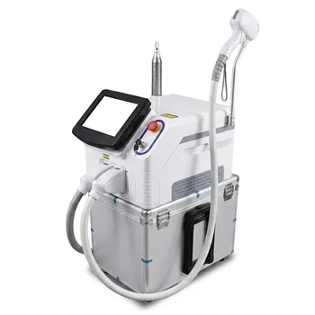
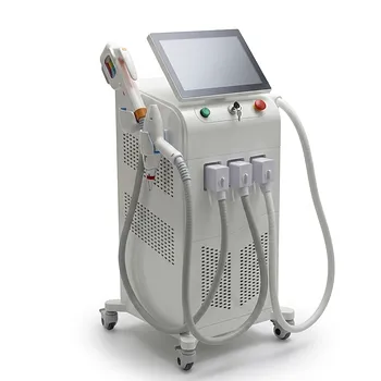
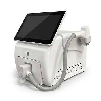
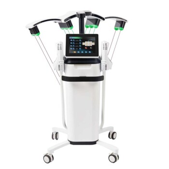
For benign acquired melanocytic nevi, laser selection should follow lesion depth and morphology—not pigment color alone. Small, superficial lesions may respond to nanosecond Q-switched lasers, including Q-switched ruby, alexandrite, or Nd:YAG systems. Deeper or thicker lesions may require long-pulsed 810 nm diode or long-pulsed alexandrite lasers, while superficial raised lesions can be treated with Er:YAG or pulsed CO₂ ablation by an experienced dermatologist.
The central principle is depth matching: Q-switched lasers target superficial or dermal melanin selectively, whereas Er:YAG, CO₂, and selected long-pulsed systems remove or thermally treat thicker tissue. These treatments should only follow a reliable diagnosis of benignity, because laser treatment can destroy diagnostic tissue and may leave residual nevus cells.
Match the Laser to the Nevus
Small and superficial nevi
For small, flat, superficially pigmented nevi, suitable technologies include nanosecond Q-switched ruby, alexandrite, and Nd:YAG lasers.
These systems deliver short, high-peak-power pulses that preferentially fragment melanin with less nonspecific thermal injury than long-pulsed devices.
Deeper or thicker lesions
For thicker lesions or lesions with deeper melanocytic nests, the primary reference identifies 810 nm diode and long-pulsed alexandrite systems as potential options.
Reported treatment parameters commonly use approximately 40–60 J/cm², 8–12 mm spot sizes, and 20–30 ms pulse durations. These settings are not universal prescriptions; they must be adjusted for lesion thickness, skin type, location, and the specific device.
Raised or papillomatous lesions
Er:YAG and pulsed CO₂ lasers can be used to ablate elevated, papillomatous lesions when the diagnosis is secure and the required depth can be controlled.
Er:YAG generally provides more superficial, precise ablation with limited thermal injury, while CO₂ provides greater coagulation and hemostasis but can produce more thermal damage.
Parameter Considerations
Fluence and pulse duration
The cited long-pulsed approach uses a fluence range of 40–60 J/cm², with pulse durations of 20–30 ms and relatively large 8–12 mm spots.
Longer pulses and sufficient fluence are intended to deliver heat deeper into the lesion, where melanocytic nests may not be reached adequately by superficial pigment targeting alone.
Spot size
Larger spots, such as 8–12 mm, can improve treatment uniformity and efficiency for suitable lesions.
A smaller spot, such as 2–4 mm, may be selected for small or anatomically confined lesions. The spot size must correspond to the lesion dimensions and the device’s optical and thermal characteristics.
Q-switched Nd:YAG example
One supplementary protocol describes a 1,064 nm Q-switched Nd:YAG laser using a 2 mm spot, 10 Hz frequency, and a stated 10–12 J output.
Because the reference does not clearly specify whether the final value represents joules per pulse, power, or another device-specific setting, it should not be treated as a transferable fluence recommendation. The operator must calculate and document the actual energy density in J/cm².
Cooling
Integrated contact epidermal cooling should be adjusted according to the patient’s Fitzpatrick skin type and treatment site.
Cooling protects the epidermis, limits unwanted thermal injury, and may reduce the risk of post-inflammatory pigment alteration and scarring. It does not eliminate the need for conservative test spots and appropriate follow-up.
When Combination Treatment Is Considered
Ablation followed by pigment targeting
For selected deeper lesions, a normal-mode or ablative laser can first remove the overlying epidermis. A Q-switched pigment laser may then be used to improve access to deeper melanocytic components.
The rationale is optical: removing the superficial layer can reduce obstruction and allow subsequent Q-switched energy to reach deeper pigment. This approach is more aggressive than Q-switched treatment alone and requires careful depth control.
Compound nevi
For compound nevi, supplementary protocols describe Er:YAG ablation of the visible superficial component, followed by either:
- One or two single-pulse CO₂ passes for additional coagulation and hemostasis, or
- A pass with Q-switched 1,064 nm Nd:YAG to target deeper pigment.
These approaches are not interchangeable. CO₂ adds thermal coagulation, whereas Nd:YAG provides selective pigment targeting.
Dermal nevi
Er:YAG and pulsed CO₂ systems may be considered for selected dermal lesions when the depth and benign diagnosis are well established.
However, deeply pigmented dermal nevi present a major diagnostic and clearance problem. Residual dermal melanocytes may remain after laser treatment, and recurrent pigmentation can resemble melanoma or a pseudomelanoma.
Why Diagnosis Must Come First
Laser is not a substitute for histology
A lesion should not be treated solely because it appears cosmetically typical. Any lesion with clinical, dermoscopic, or historical atypia should undergo appropriate dermatologic assessment and, when indicated, biopsy.
Laser destruction can remove the tissue needed to confirm the diagnosis and may complicate evaluation of recurrence.
Pigmented dermal lesions require special caution
The supplementary material appropriately highlights that laser treatment of pigmented dermal nevi can leave residual pigmented cells.
The statement that such lesions are “strictly contraindicated” for all laser treatment is too broad as a general rule. The defensible clinical conclusion is narrower: laser treatment should be avoided when benignity is uncertain or when complete histologic clearance is important, and treatment of confirmed lesions should be undertaken only by specialists who understand the risk of recurrence and diagnostic confusion.
Do not confuse nevi with Hori’s macules
Acquired bilateral nevus of Ota-like macules, or Hori’s macules, are dermal hyperpigmentation disorders rather than ordinary acquired melanocytic nevi.
They are commonly treated with Q-switched devices, particularly 1,064 nm Nd:YAG, and sometimes combined wavelength protocols. Their treatment parameters and clinical objectives should not be directly transferred to benign acquired nevi.
Understanding the Trade-offs
Q-switched lasers alone may be incomplete
Q-switched lasers can provide good pigment reduction in superficial lesions, but intact epidermis and lesion depth may limit energy delivery to deeper nevus cell nests.
Clinical fading therefore does not necessarily mean complete histologic removal.
Ablative lasers can scar or alter pigmentation
Er:YAG and CO₂ lasers provide direct tissue removal but carry risks of prolonged erythema, infection, scarring, and post-inflammatory hyperpigmentation or hypopigmentation.
These risks increase with excessive depth, repeated passes, aggressive fluence, and treatment of darker skin types.
Higher energy is not automatically better
The cited ranges of 25 J/cm² in one ablative context and 40–60 J/cm² in a long-pulsed context should not be combined or generalized across devices.
Fluence, spot size, pulse duration, repetition rate, cooling, and tissue response are interdependent. A numerical setting is meaningful only in relation to the specific laser platform and handpiece.
Cosmetic clearance is not the same as cure
Laser treatment may improve visible pigmentation while leaving viable melanocytic nests behind.
Patients should be informed about recurrence, incomplete clearance, pigmentary changes, and the possibility that recurrent pigmentation may require biopsy.
How to Apply This to Your Project
The safest protocol is one built around confirmed diagnosis, lesion depth, device-specific calibration, conservative test treatment, and documented follow-up.
- If your primary focus is superficial, flat nevi: Consider a nanosecond Q-switched ruby, alexandrite, or Nd:YAG platform, using conservative device-validated settings rather than assuming the long-pulsed 40–60 J/cm² range applies.
- If your primary focus is deeper or thicker nevi: Evaluate long-pulsed 810 nm diode or alexandrite systems, with the cited starting framework of 40–60 J/cm², 8–12 mm spots, and 20–30 ms pulses only under specialist supervision.
- If your primary focus is raised or papillomatous lesions: Consider precisely controlled Er:YAG or pulsed CO₂ ablation after confirming benignity and planning the intended ablation depth.
- If your primary focus is compound or dermal lesions: Use a specialist-led staged or combination approach only when the diagnosis is secure, recognizing that visible clearance may not equal histologic clearance.
- If your primary focus is patient safety: Prioritize dermatologic examination, dermoscopy or biopsy when indicated, skin-type-adjusted cooling, test spots, and long-term surveillance.
The right laser is the one that matches the lesion’s depth while preserving diagnostic safety and minimizing unnecessary tissue injury.
Summary Table:
| Nevus Type | Recommended Laser | Key Parameters | Considerations |
|---|---|---|---|
| Small, superficial | Q-switched ruby, alexandrite, Nd:YAG | Short pulses; fluence per device | Preferentially target melanin; minimal thermal injury |
| Deeper or thicker | Long-pulsed 810nm diode, alexandrite | 40–60 J/cm², 20–30 ms, 8–12 mm spot | Heat deeper nests; adjust for skin type and lesion |
| Raised or papillomatous | Er:YAG, pulsed CO₂ | Ablative fluence per device | Precise ablation; CO₂ offers hemostasis, more thermal damage |
| Compound or dermal | Combination (ablation + Q-switched) | Staged, specialist-led | Residual nests; risk of pseudomelanoma |
Ensure safe, effective treatments with BELIS's advanced laser platforms. From Q-switched to long-pulsed technologies, our professional equipment is designed for clinics and premium salons. Contact us today to find the perfect laser system for your practice and elevate patient outcomes.
Related Products
- 808nm Diode Laser Hair Removal Machine and Equipment with Picolaser Arm
- Hydrofacial Machine with Facial Skin Analyzer and Skin Tester
- 808nm Diode Laser Hair Removal Machine 755+808+1064nm Mixed Wavelength Professional Equipment
- Trilaser Diode Hair Removal Machine for Beauty Clinic Use
- Clinic Use IPL SHR ND YAG Laser Hair Removal RF Skin Tightening Machine
People Also Ask
- How do 800/810 nm diode hair removal lasers compare to 1064 nm long-pulsed Nd:YAG lasers? Discover the Best for Your Patients
- What ocular safety protocols are required when operating diode or alexandrite hair removal lasers near the periorbital area? Essential eye protection and shielding for safe treatment.
- How do 800–810 nm Diode hair removal lasers compare to 1064 nm Long-Pulsed Nd:YAG lasers in terms of hair reduction efficacy and patient comfort? Discover Key Differences for Optimal Hair Removal
- How do broad-spectrum noncoherent light sources compare to single-wavelength diode lasers in aesthetic hair removal applications? Find the best fit for your clinic.
- How can aesthetic practitioners prevent side effects like paradoxical hair regrowth and thermal burns when performing diode laser hair removal on dark skin? Master safe protocols for Fitzpatrick IV–VI skin.