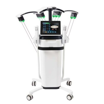
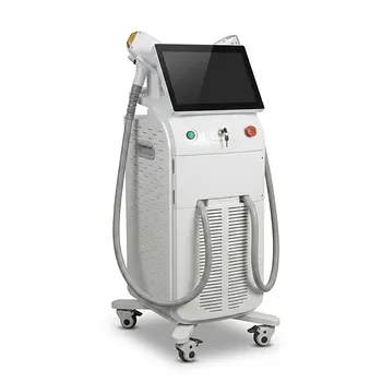
Fractional laser photothermolysis works by delivering laser energy in a microscopic, patterned array rather than treating the entire skin surface. Wavelength-specific absorption, primarily by tissue water, converts light into confined columns of thermal injury called microthermal treatment zones (MTZs). The resulting wound-healing response promotes epidermal repair and dermal collagen remodeling, while patient factors such as pigmentation, skin thickness, sun exposure, age, sex, and healing history determine how much energy and treatment density the skin can safely tolerate.
The central principle is controlled injury with preserved recovery capacity: fractional treatment creates enough thermal damage to stimulate remodeling, but leaves adjacent viable tissue available to support repair. Dosimetry must therefore balance the intended depth and remodeling effect against the patient’s individual risk of pigmentary change, prolonged inflammation, and thermal injury.
How Fractional Photothermolysis Produces Its Effect
Selective Absorption Creates Thermal Injury
Fractional systems deliver energy at specific wavelengths, including 10,600 nm carbon dioxide and fractional erbium wavelengths. These wavelengths are strongly absorbed by water in the skin, converting optical energy into heat within a targeted microscopic volume.
The goal is not generalized heating. It is spatially confined photothermal injury that reaches a selected depth while limiting damage outside the treatment columns.
Microthermal Zones Control the Injury Pattern
The laser divides treatment into an array of microscopic columns rather than one continuous ablated or coagulated field. These MTZs may extend from the epidermis into the dermis, depending on wavelength, pulse characteristics, and delivered energy.
The untreated tissue between zones remains viable. It supplies keratinocytes, fibroblasts, signaling molecules, and other cellular resources that support epithelial reconstruction and wound repair.
The Wound-Healing Cascade Drives Remodeling
Thermal injury initiates a controlled inflammatory and reparative response. Heat-shock proteins, including HSP70, participate in cellular stress signaling, while fibroblast activity contributes to new extracellular matrix production.
The remodeling response includes increased synthesis and reorganization of collagen, including early type III collagen, followed by longer-term dermal restructuring. This can improve fine lines, acne scars, and features of photodamage.
Fractionation Preserves a Biological Reservoir
Fractional treatment depends on the presence of surrounding healthy tissue. This intact tissue acts as a local reservoir for cells and signaling factors, allowing treated areas to repair more rapidly than a fully resurfaced skin surface.
That principle explains the typical clinical trade-off: fractional treatment can produce meaningful remodeling with less downtime than fully ablative resurfacing, although the magnitude of benefit depends on treatment intensity and the condition being treated.
Which Dosimetry Variables Matter
Fluence Determines the Energy Delivered
Fluence is the energy delivered per unit area. Increasing fluence generally increases the thermal effect and can increase treatment depth or severity, depending on the device and settings.
Higher fluence may improve the treatment effect in selected cases, but it also raises the risk of excessive coagulation, ablation, prolonged inflammation, and pigmentary complications.
Density Determines How Much Skin Is Treated
Treatment density describes how many MTZs are created within a given area. A higher density means a greater proportion of the skin is injured during one pass or treatment session.
Density must be considered separately from energy per MTZ. A moderate energy delivered at high density can create substantial cumulative injury, while a higher energy delivered at low density may produce deeper but more widely spaced zones.
Pulse Duration Shapes Thermal Confinement
Pulse duration affects how heat accumulates and dissipates. A pulse that is appropriately matched to the target tissue and treatment objective can confine thermal injury more effectively.
The practical goal is to create the intended MTZ without allowing heat to spread excessively into adjacent tissue. Device-specific pulse behavior means settings cannot be transferred reliably between platforms solely by comparing numerical values.
Depth and Wavelength Define the Target
Wavelength influences how energy interacts with water and therefore where thermal energy is deposited. Carbon dioxide and erbium systems can differ in ablation, coagulation, penetration, and recovery characteristics.
Dosimetry should match the intended target: superficial epidermal correction, deeper dermal remodeling, scar treatment, or a combination of objectives. The same setting is not appropriate for every indication or anatomical site.
Patient Factors That Change Safe Dosimetry
Baseline Pigmentation Affects Risk
Skin with greater baseline melanin content has a higher risk of post-inflammatory hyperpigmentation after laser-induced inflammation. This risk is particularly relevant when treating darker skin types or patients with a history of pigmentary change after injury.
Pigmentation does not automatically exclude treatment, but it generally demands more conservative energy or density, careful patient selection, and appropriate peri-treatment management.
Recent Sun Exposure Increases Reactivity
Recent tanning or ultraviolet exposure can increase epidermal melanin activity and skin reactivity. It may also make post-treatment pigmentary changes more likely.
Treatment planning should account for recent sun exposure rather than relying only on the patient’s usual skin type. Elective treatment may need to be deferred until the skin has returned to a stable baseline.
Tissue Thickness Changes the Thermal Target
Skin thickness varies by anatomical location, age, sex, and individual structure. Thin skin provides less tissue margin between the treatment surface and deeper structures, while thicker or more fibrotic tissue may require a different strategy to achieve the intended remodeling.
Periocular skin, the neck, and other delicate sites should not automatically receive the same dosimetry used on thicker facial areas.
Age Influences Repair Capacity
Age affects epidermal turnover, dermal thickness, collagen production, vascularity, and the speed of wound repair. Older skin may have a reduced capacity for rapid recovery and may respond differently to the same thermal dose.
Age should therefore inform treatment intensity and recovery expectations, but it should not be used as a standalone predictor. Overall skin condition and medical history remain important.
Sex and Hormonal Context May Matter
Sex-associated differences in skin thickness, sebaceous activity, hair-bearing areas, and hormonal environment can influence tissue response and healing. These differences are typically considered alongside anatomy and clinical indication rather than used as independent dosing rules.
The practical implication is that dosimetry should be individualized rather than standardized solely by age or sex.
Personal Healing History Is Clinically Important
A history of prolonged redness, abnormal scarring, post-inflammatory hyperpigmentation, or delayed healing provides useful information about the patient’s response to controlled injury.
Patients with impaired healing, active infection, or a tendency toward abnormal scarring require particularly careful assessment. The treatment plan should reflect both the desired result and the patient’s demonstrated capacity to recover.
Hydration and Skin Condition Affect Response
The water content and condition of the epidermis influence how energy is absorbed. Irritated, inflamed, recently exfoliated, or otherwise compromised skin may respond less predictably and may have a higher risk of excessive inflammation.
The treatment area should be evaluated in its current condition, not only by its diagnosis or the intended procedure.
Why the Same Settings Do Not Suit Every Patient
Dosimetry Is a Biological, Not Merely Technical, Decision
Laser parameters describe the energy delivered by the device, but the clinical effect depends on how the patient’s tissue absorbs and responds to that energy. Two patients treated with identical settings can develop different MTZ severity, inflammation, recovery time, and pigmentary outcomes.
For this reason, dosimetry should combine device parameters with patient-specific assessment and anatomical judgment.
Treatment Area Changes the Margin for Error
Facial subunits differ in thickness, vascularity, sebaceous activity, and exposure to movement or friction. Areas near the eyes, lips, and jawline may require different settings from the forehead or cheeks.
A uniform full-face setting can therefore create uneven risk and uneven results. Treatment planning should account for regional anatomy.
Test Areas and Response-Based Adjustment Can Improve Control
When appropriate, a clinician may use a test area or begin with conservative parameters and assess the immediate tissue response. Subsequent treatment can then be adjusted according to erythema, edema, thermal endpoint, recovery history, and clinical goals.
This approach is especially relevant when pigmentary risk or the patient’s healing response is uncertain.
Understanding the Trade-offs
More Energy Is Not Automatically More Effective
Increasing fluence can increase thermal remodeling, but it also increases the probability of excessive injury. The desired endpoint is effective stimulation of repair, not maximal tissue destruction.
Overtreatment can produce prolonged erythema, edema, burns, delayed epithelial recovery, infection risk, or persistent pigmentary change.
Higher Density Increases Cumulative Injury
Increasing density treats more of the skin during the same session. Although this may improve uniformity or accelerate correction, it reduces the amount of untreated tissue available to support immediate recovery.
High density can therefore be inappropriate when the patient has darker pigmentation, recent sun exposure, thin skin, or a history of prolonged inflammation.
Fractional Treatment Still Has Meaningful Risks
Fractionation reduces the treated surface area, but it does not eliminate thermal injury. Both ablative and nonablative fractional procedures can cause pain, swelling, erythema, infection, pigmentary alteration, and, less commonly, scarring.
Shorter downtime compared with fully ablative resurfacing should not be interpreted as zero downtime or zero risk.
Recovery Time Can Be Misleading
Fractional treatments often produce approximately one to two days of transient redness and mild edema, but recovery varies with wavelength, fluence, density, treatment area, and patient biology.
A patient’s visible recovery may be brief while deeper remodeling continues for considerably longer. Clinical expectations should distinguish immediate recovery from the time required for collagen reorganization.
Making the Right Choice for Your Goal
The most defensible plan is the one that matches treatment intensity to both the clinical target and the patient’s recovery and pigmentary risk.
- If your primary focus is deeper dermal remodeling: Use a treatment strategy that reaches the intended dermal target while controlling fluence, pulse behavior, and density to avoid excessive thermal spread.
- If your primary focus is minimizing downtime: Favor a fractional approach with conservative-to-moderate treatment coverage, recognizing that lower density or energy may require multiple sessions.
- If your primary focus is reducing pigmentary complications: Give particular weight to baseline pigmentation, recent sun exposure, prior hyperpigmentation, and conservative density selection.
- If your primary focus is treating thin or sensitive skin: Adjust parameters for regional tissue thickness and healing capacity rather than applying a uniform full-face setting.
- If your primary focus is predictable recovery: Incorporate the patient’s prior wound-healing response and use clinical endpoints or staged treatment to guide intensity.
Successful fractional photothermolysis depends on creating enough controlled injury to stimulate remodeling while preserving enough viable tissue for reliable repair.
Summary Table:
| Mechanism/Patient Factor | Description | Clinical Relevance |
|---|---|---|
| Mechanisms | ||
| Selective Absorption | Wavelength-specific water absorption converts light to heat | Determines depth and severity of thermal injury |
| Microthermal Zones (MTZs) | Patterned columns of thermal injury with viable surrounding tissue | Balances remodeling with recovery capacity |
| Wound-Healing Cascade | HSP70, fibroblast activation, collagen remodeling | Drives long-term skin improvement |
| Biological Reservoir | Untreated tissue provides cells for repair | Enables faster recovery vs full ablation |
| Dosimetry Variables | ||
| Fluence | Energy per unit area | Higher fluence increases thermal effect and risk |
| Density | MTZs per area | Higher density increases cumulative injury |
| Pulse Duration | Influences thermal confinement | Affects MTZ shape and surrounding damage |
| Wavelength | Determines water absorption and depth | Matches target indication and anatomical site |
| Patient Factors | ||
| Pigmentation | Baseline melanin content | Higher risk of post-inflammatory hyperpigmentation |
| Sun Exposure | Recent UV exposure increases reactivity | May require treatment delay or conservative settings |
| Tissue Thickness | Varies by site, age, sex | Affects safety margins and target depth |
| Age | Influences repair capacity | Adjust intensity and recovery expectations |
| Sex & Hormonal Context | Skin thickness and healing differences | Individualize dosing rather than use standalone rules |
| Healing History | Prior responses to injury | Identifies abnormal scarring or hyperpigmentation risk |
| Hydration & Skin Condition | Water content and irritation | Affects absorption and predictability of response |
| Trade-offs | ||
| More Energy ≠ More Effective | Increased risk of complications | Balance stimulation with safety |
| Higher Density Increases Injury | Less viable tissue for repair | Use cautiously in high-risk patients |
| Fractional Still Has Risks | Pain, swelling, infection, scarring | Not zero downtime or zero risk |
| Recovery vs Remodeling | Visible recovery vs deep collagen changes | Set realistic expectations for results duration |
Ready to elevate your aesthetic practice with advanced fractional laser technology? At BELIS, we specialize in professional-grade medical aesthetic equipment exclusively for clinics and premium salons. Our portfolio includes cutting-edge fractional lasers (CO2, Erbium) designed for precise photothermolysis and optimal patient outcomes. By partnering with us, you gain access to:
- Advanced Technology: State-of-the-art laser systems with customizable parameters for individualized treatment.
- Comprehensive Support: OEM/ODM services, full certifications, and reliable supply chain.
- Expert Guidance: Our team helps you integrate safe and effective dosimetry protocols into your practice.
Ensure safe and effective treatments for your patients while maximizing your practice's potential. Contact us today to discuss how our solutions can meet your needs.
Related Products
- Fractional CO2 Laser Machine for Skin Treatment
- Fractional CO2 Laser Machine for Skin Treatment
- RF Microneedling Machine Micro Needle Radio Frequency Machine
- RF Microneedling Machine Micro Needle Radio Frequency Machine
- Ultrasonic Cavitation Machine Lipo Laser Device
People Also Ask
- What is the technical principle behind CO2 Laser Fractional micro-perforations? Master Scar Revision Mechanics
- What parameters and treatment intervals are advised when applying fractional CO2 laser technology to delicate periorbital skin laxity? Discover safe protocols for eyelid rejuvenation.
- How do energy-based modalities complement injectable neurotoxins in décolleté rejuvenation? Explore synergistic benefits.
- What is the primary function of a high-precision fractional CO2 laser system for GSM? Restore Vaginal Health Naturally
- Why do fractional CO2 laser parameters need to be differentiated? Master Keloid vs. Hypertrophic Scar Treatment