For deep, nodular, or exophytic vascular lesions, 1064 nm Nd:YAG is chosen because it reaches larger and deeper vascular channels that shorter visible wavelengths may not adequately treat. A long-pulsed 1064 nm treatment is configured with a small, lesion-matched spot, carefully selected pulse duration and fluence, continuous epidermal cooling, and a conservative endpoint of slight immediate shrinkage or light bluing. A sequential visible-light/1064 nm approach can enhance absorption in blood, potentially allowing lower visible-light fluence while extending treatment effect into deeper vessels.
The central principle is depth-controlled thermal coagulation without exceeding the minimum effective dose. Parameters must be individualized to lesion thickness, vessel caliber, skin type, device architecture, and cooling performance; published ranges are not interchangeable across laser systems.
Why 1064 nm Is Used for Nodular Lesions
Deeper optical penetration
The 1064 nm wavelength penetrates substantially deeper than green or yellow vascular wavelengths. This makes it useful for thickened, exophytic, nodular, or mature vascular structures whose clinically important vessels lie beyond the effective reach of superficial light.
The wavelength is also relatively less absorbed by epidermal melanin than shorter wavelengths, providing a useful safety advantage in darker skin phototypes. However, the higher fluences commonly required still create a meaningful risk of epidermal and dermal thermal injury.
Treatment of larger-caliber vessels
Hemoglobin absorption is lower at 1064 nm than at green or yellow wavelengths. The trade-off is that the laser generally requires greater delivered energy to produce coagulation in deeper or larger vessels.
This is why 1064 nm is particularly relevant to resistant vascular malformations, deep venous components, and nodular lesions rather than as a default choice for every superficial vascular lesion.
Why nodules require conservative dosing
A nodule contains a greater volume of vascular and nonvascular tissue than a flat telangiectatic vessel. Excessive energy can therefore create a large zone of thermal injury, increasing the risk of necrosis, ulceration, scarring, and post-inflammatory hyperpigmentation.
The goal is not dramatic immediate ablation. It is controlled coagulation and progressive involution with the lowest effective thermal burden.
How the 1064 nm Treatment Is Configured
Spot size
For exophytic nodules, the spot should generally be small and closely matched to the target diameter. Reported approaches use approximately 2–4 mm spots, including a 3 mm spot for selected deeper nodules such as glomus tumors or blue rubber bleb nevi.
A smaller spot improves targeting of a discrete nodule but increases the risk of excessive fluence density if the energy is not adjusted appropriately. The operator should avoid unintended overlap unless the specific device protocol explicitly validates it.
Pulse duration
Pulse duration should reflect the vascular caliber and target depth. Published clinical approaches vary substantially:
- Some long-pulsed systems use approximately 3–15 ms for selected vascular applications.
- Other protocols begin around 30–40 ms for thicker or larger-caliber vessels.
- A nodule-specific approach cited in the primary reference uses approximately 50 ms.
These values should not be treated as universal prescriptions. Pulse width, spot size, fluence, cooling, and the device’s actual pulse profile interact; changing one variable may require recalibration of the others.
Fluence
For selected deep nodular lesions, the cited treatment range is approximately 80–200 J/cm² with a pulse width near 50 ms, using a 2–4 mm spot. This is a broad range, not a routine starting setting.
Other 1064 nm vascular protocols report lower or different fluence ranges depending on lesion color, vessel depth, pulse duration, and device design. Therefore, the appropriate setting is the lowest fluence that produces the desired clinical endpoint, rather than a fixed number selected solely from lesion diagnosis.
Cooling
Integrated contact or surface cooling is essential. Cooling may be provided through a cooled handpiece, chilled coupling medium, or another validated pre-, intra-, and post-pulse method.
Cooling protects the epidermis while allowing energy to reach deeper tissue. It does not eliminate the risk of deep thermal damage, particularly when treating bulky nodules or when pulses overlap.
Pulse pattern
Use a non-overlapping pattern unless a validated protocol specifically requires otherwise. Pulse stacking with long-pulsed Nd:YAG treatment should generally be avoided because residual heat can accumulate and produce scarring or textural change.
For deeper lesions, treatment may require staged sessions rather than attempting complete regression in a single pass.
Clinical Endpoint and Safety Limits
Desired endpoint
For nodular or exophytic lesions, the preferred endpoint is slight immediate shrinkage, mild flattening, or light bluing. These signs suggest that vascular heating has occurred without overwhelming tissue injury.
The endpoint should be assessed across the entire lesion, including its base and any clinically deeper component.
Endpoints indicating overtreatment
Deep gray discoloration, marked whitening, blistering, tissue charring, or complete immediate collapse should be treated as warning signs rather than therapeutic success. They indicate excessive thermal injury or energy concentration.
Vessel rupture and pronounced purpura are also undesirable when the objective is controlled coagulation rather than mechanical vessel disruption.
Anesthesia and monitoring
Thicker nodules may require local anesthesia because the treatment deposits substantial energy into deeper tissue. Anesthesia can also reduce pain-driven movement, but it may remove useful feedback; visual endpoints and conservative titration therefore become especially important.
The lesion should be reassessed after treatment and during follow-up for delayed necrosis, ulceration, pigmentary alteration, and scarring.
When a Dual-Wavelength Approach Is Rational
Sequential optical absorption
A dual-wavelength approach typically delivers a visible green or yellow pulse first, followed by a 1064 nm pulse. The visible wavelength heats blood and promotes conversion of oxyhemoglobin to methemoglobin.
Methemoglobin absorbs near-infrared energy more effectively than the original blood state. When the 1064 nm pulse follows after a short delay, the altered optical state can increase deep energy deposition without requiring the same visible-light fluence that would otherwise be necessary.
Timing between pulses
The primary reference describes a delay of approximately 10–50 ms between the visible pulse and the 1064 nm pulse. The correct delay is device- and protocol-dependent and should not be extrapolated without confirming the system’s pulse sequencing and thermal behavior.
The purpose of the delay is to permit the intended blood-heating and absorption shift while limiting unnecessary epidermal heat accumulation.
Why this may help nodular lesions
The sequential approach can address different vascular depths in the same lesion: the first wavelength treats more superficial components, while the second benefits from deeper penetration and the altered blood absorption profile.
This may be useful for lesions with a superficial red component overlying a deeper blue, violaceous, or nodular component. It is not automatically superior to 1064 nm alone, particularly when the main target is very deep or when the superficial component is minimal.
Understanding the Trade-offs
Higher penetration means higher consequence
The same penetration that makes 1064 nm useful for nodules also increases the possibility of injury to deeper dermis and subcutaneous tissue. A visually modest surface response does not necessarily mean that the deeper treatment effect is modest.
This is why aggressive escalation based only on surface appearance is unsafe.
Published parameters vary
Fluence and pulse-width ranges differ across systems because spot size, beam profile, pulse shape, cooling technology, and calibration differ. A setting that is appropriate on one platform cannot be copied directly to another.
The ranges above should therefore be interpreted as reported clinical starting frameworks, not universal standards of care.
Dual wavelength is not risk-free
The visible pulse may reduce the fluence needed for the subsequent 1064 nm pulse, but it also adds heat to the treatment sequence. The combined exposure must be assessed as one thermal event.
Cooling, pulse spacing, non-overlap, and conservative endpoint assessment remain necessary.
Disease diagnosis still matters
“Nodular vascular lesion” is a treatment morphology, not a complete diagnosis. Glomus tumors, blue rubber bleb nevi, vascular malformations, Kaposi sarcoma nodules, and other lesions differ in depth, blood flow, tissue composition, and recurrence behavior.
A lesion that is atypical, rapidly changing, ulcerated, painful, or diagnostically uncertain should be evaluated appropriately before laser treatment. Laser coagulation does not substitute for diagnosis or oncologic management.
How to Apply This to the Clinical Goal
Parameter selection should be performed by a trained clinician using the specific device’s validated protocol, test areas, cooling system, and patient-specific risk assessment.
- If your primary focus is a discrete exophytic or nodular target: Use a small, lesion-matched spot—often in the 2–4 mm range—with conservative titration toward slight shrinkage or light bluing rather than complete immediate collapse.
- If your primary focus is a deep, thick vascular component: Favor 1064 nm because of its penetration, and adjust pulse duration and fluence to vessel caliber and depth while maintaining continuous cooling.
- If your primary focus is mixed superficial and deep vascular involvement: Consider a visible-light pulse followed approximately 10–50 ms later by 1064 nm when the platform supports validated sequential delivery.
- If your primary focus is minimizing scarring: Avoid pulse stacking and overlapping pulses, use the lowest effective fluence, and stop when gray discoloration, excessive whitening, blistering, or other overtreatment signs appear.
- If your primary focus is treating an uncertain or tumor-like nodule: Establish the diagnosis and management plan before laser coagulation, particularly for lesions with atypical growth, ulceration, or suspected malignancy.
The safest and most effective approach is to match wavelength, pulse duration, fluence, spot size, cooling, and endpoint to the lesion’s actual vascular depth—not simply to its surface color.
Summary Table:
| Parameter | Recommended Setting | Clinical Rationale |
|---|---|---|
| Wavelength | 1064 nm (Nd:YAG) | Deeper penetration for nodular/exophytic lesions; lower melanin absorption for darker skin types. |
| Spot Size | 2–4 mm | Matches lesion diameter; reduces risk of overtreatment and allows precise targeting. |
| Pulse Duration | 3–50 ms (varies by system) | Matches vessel caliber and depth; longer pulses for larger/deeper vessels. |
| Fluence | 80–200 J/cm² (for 50 ms pulse) | Lowest effective dose to achieve slight shrinkage; avoid overtreatment. |
| Cooling | Continuous contact cooling | Protects epidermis, reduces thermal injury risk. |
| Pulse Pattern | Non-overlapping | Avoids heat accumulation and scarring. |
| Endpoint | Slight shrinkage, light bluing | Indicates controlled coagulation without excessive damage. |
| Dual-Wavelength | Visible light + 1064 nm (delay 10–50 ms) | Enhances methemoglobin absorption for deeper effect; useful for mixed superficial/deep lesions. |
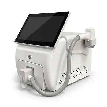
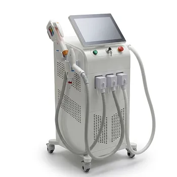
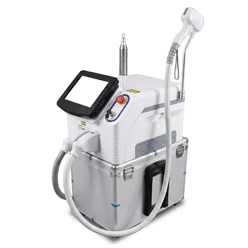
Ready to elevate your clinic's vascular treatment capabilities? BELIS offers advanced aesthetic laser systems, including 1064 nm Nd:YAG and dual-wavelength platforms, designed for safe and effective treatment of even the most challenging vascular lesions. Our equipment is trusted by clinics and premium salons worldwide. Contact us today to learn how our technology can expand your service offerings and improve patient outcomes. With BELIS, you get professional-grade equipment, comprehensive support, and solutions tailored to your practice.
Related Products
- Tri Laser Diode Hair Removal Machine Professional Beauty Equipment
- 808nm Diode Laser Hair Removal Machine 755+808+1064nm Mixed Wavelength Professional Equipment
- Q Switch Nd Yag Laser Machine Tattoo Removal Nd Yag Machine
- Clinic Use IPL and SHR Hair Removal Machine with Nd Yag Laser Tattoo Removal
- Clinic Use IPL SHR ND YAG Laser Hair Removal RF Skin Tightening Machine
People Also Ask
- How do 800/810 nm diode hair removal lasers compare to 1064 nm long-pulsed Nd:YAG lasers? Discover the Best for Your Patients
- What is the recommended treatment interval for diode laser hair removal? Achieve Max Hair Reduction with 45-Day Sessions
- How do broad-spectrum noncoherent light sources compare to single-wavelength diode lasers in aesthetic hair removal applications? Find the best fit for your clinic.
- How can aesthetic practitioners prevent side effects like paradoxical hair regrowth and thermal burns when performing diode laser hair removal on dark skin? Master safe protocols for Fitzpatrick IV–VI skin.
- What long-term hair reduction results can aesthetic clinics expect when utilizing diode laser hair removal devices? Achieve Durable Outcomes with Proper Protocols