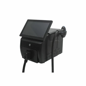
For semi-selective coagulation of verrucous epidermal nevi, begin conservatively with approximately 2 W, a 0.5 ms pulse duration, and a 2–3 mm spot size. The desired endpoint is uniform white-gray blanching of the treated tissue, without carbonization, charring, or excessive surface injury. Large lesions should be treated using an interrupted stripe or polka-dot pattern, preceded by a test spot and a 3–6 month observation period before extensive treatment.
The procedure is guided more reliably by the tissue endpoint than by a fixed energy setting: achieve homogeneous white-gray coagulation while avoiding charring, and limit contiguous treatment to reduce scarring and contracture.
Establishing the Treatment Parameters
Start With Conservative Baseline Settings
A reasonable initial setting for a 532-nm Nd:YAG system is:
- Power: approximately 2 W
- Pulse duration: approximately 0.5 ms
- Spot size: 2–3 mm
Some systems and treatment protocols use a 2–4 mm spot size. The correct setting depends on the device’s pulse architecture, beam profile, handpiece, lesion thickness, and tissue response.
Adjust for the Specific Laser System
Power and pulse duration should not be transferred directly between different laser platforms. A Q-switched device, a millisecond long-pulse device, and a system displaying fluence rather than power require different parameter frameworks.
Fluence values cited for treating pigmented epidermal lesions should not automatically be applied to verrucous epidermal nevi. Those protocols target melanin and different clinical endpoints, whereas semi-selective coagulation of a verrucous nevus is primarily guided by controlled thermal tissue change.
Use the Lowest Effective Exposure
Treatment should begin at a level that produces the intended blanching without epidermal disruption beyond the treatment target. Incremental adjustment may be considered when the endpoint is not reached, but excessive energy, overlap, or pulse stacking increases the risk of blistering, ulceration, dyspigmentation, and scarring.
Recognizing the Correct Clinical Endpoint
Target Homogeneous White-Gray Blanching
The preferred endpoint is even white-gray discoloration across the treated tissue. This indicates the intended degree of thermal coagulation for the semi-selective technique.
The endpoint should be assessed across the treatment field rather than inferred solely from the console setting.
Avoid Carbonization and Charring
Blackening, carbonization, smoke, or obvious surface charring indicates excessive thermal injury. Additional irradiation should be stopped in that area, and the parameters should be reassessed before proceeding.
Charring is not evidence of a better treatment effect. It increases surface damage and may increase the likelihood of prolonged healing and scar formation.
Monitor for Excessive Thermal Accumulation
Avoid unnecessary pulse overlap and allow sufficient assessment between treatment passes. Particular caution is warranted in darker skin types because 532-nm light is strongly absorbed by epidermal melanin, increasing the risk of nonspecific epidermal injury and post-inflammatory pigmentary change.
Selecting the Treatment Technique
Use Test Spots Before Treating Extensive Areas
A test spot should be treated first, followed by observation for approximately 3–6 months. This interval helps assess the patient’s scar response, pigmentary response, healing behavior, and early recurrence.
A favorable test response supports proceeding with a larger treatment area, but it does not eliminate the need for conservative coverage and follow-up.
Use Stripe or Polka-Dot Coverage
Large-surface lesions should be treated with an interrupted pattern, such as:
- Stripe or “zebra-stripe” treatment
- Polka-dot or checkerboard treatment
These approaches leave untreated bridges of tissue between coagulated zones. The spacing reduces contiguous thermal damage and helps limit contractile scarring.
Treat Large Lesions Over Multiple Sessions
Extensive verrucous nevi should generally be divided across multiple sessions rather than treated as one continuous field. Staged treatment permits evaluation of healing and scar behavior before additional tissue is exposed.
Full-field coagulation is particularly undesirable when a large lesion is located in an area where contraction could impair function or produce a conspicuous scar.
Matching the Technique to Lesion Characteristics
Soft, Papillomatous Lesions
Soft or papillomatous epidermal nevi are the more appropriate candidates for this semiselective coagulation approach. They may respond more predictably to controlled thermal coagulation than densely keratotic lesions.
The lesion’s thickness and surface architecture should still be evaluated clinically before selecting the treatment plan.
Hard, Keratotic Lesions
Hard or heavily keratotic lesions may respond poorly to semiselective laser coagulation. The laser energy may not distribute adequately through the thickened tissue, increasing the temptation to raise exposure and thereby increasing injury to surrounding or underlying skin.
Poor response should prompt diagnostic and therapeutic reassessment rather than repeated escalation without a clear endpoint.
Understanding the Trade-offs
The Endpoint Is Not a Universal Dose
A setting such as 2 W and 0.5 ms is a starting point, not a guaranteed prescription. Spot size, pulse delivery, contact technique, tissue hydration, lesion thickness, and device calibration can all change the delivered thermal effect.
For this reason, the observed tissue response should control adjustment decisions.
More Complete Coverage Can Increase Scarring
Treating every part of a large lesion in a single continuous field may appear efficient, but it increases the risk of contractile scar formation. Interrupted patterns and staged sessions trade speed for better control of thermal injury.
This trade-off is especially important near joints, eyelids, lips, or other anatomically sensitive sites.
Recurrence Remains Possible
Even when the visible lesion improves, recurrence can occur. The recommended observation period after a test spot is therefore part of treatment planning, not merely a follow-up formality.
Patients should be counseled that additional treatment may be required and that the final result must be judged after healing and adequate observation.
Pigmentary Complications Require Caution
Because 532-nm light is strongly absorbed by melanin, darker skin types may be more susceptible to blistering, persistent hypopigmentation, or post-inflammatory hyperpigmentation. Conservative test treatment, strict endpoint monitoring, and careful avoidance of excess overlap are essential.
How to Apply This to a Treatment Plan
Begin with a qualified clinical assessment confirming the diagnosis and determining whether the lesion’s morphology is suitable for semiselective coagulation. Then use the following principles:
- If your primary focus is minimizing scarring: Start near 2 W with a 0.5 ms pulse and 2–3 mm spot, use stripe or polka-dot coverage, and avoid full-field treatment.
- If your primary focus is confirming individual response: Treat a small test spot and observe it for 3–6 months before treating a broad area.
- If your primary focus is achieving controlled coagulation: Stop when homogeneous white-gray blanching appears and avoid carbonization, charring, pulse stacking, and excessive spot overlap.
- If your primary focus is treating a thick or hard keratotic lesion: Reassess suitability because these lesions may respond poorly and should not be managed by uncritical energy escalation.
A conservative, endpoint-guided, staged technique is the most defensible way to balance lesion clearance against thermal injury and scarring risk.
Summary Table:
| Parameter/Technique | Recommended Setting/Approach |
|---|---|
| Initial Power | ~2 W |
| Pulse Duration | ~0.5 ms |
| Spot Size | 2–3 mm |
| Clinical Endpoint | Uniform white-gray blanching, no charring |
| Test Spot | 3–6 months observation |
| Large Lesions | Stripe or polka-dot pattern, multiple sessions |
Maximize Treatment Success with BELIS Precision Laser Systems
At BELIS, our advanced Nd:YAG and other aesthetic lasers are designed to deliver precise, controlled outcomes for verrucous epidermal nevi and a wide range of skin concerns. Our systems feature customizable parameters, superior beam uniformity, and built-in safety features to help you achieve the desired blanching endpoint with minimal risk. Whether you're a clinic or premium salon, our equipment enhances treatment efficacy and patient safety. Explore our range of professional laser solutions today—contact us for expert guidance and to find the perfect system for your practice!
Related Products
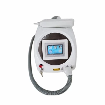
- Q Switch Nd Yag Laser Machine Tattoo Removal Nd Yag Machine
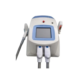
- Clinic Use IPL and SHR Hair Removal Machine with Nd Yag Laser Tattoo Removal
- Clinic Use IPL SHR ND YAG Laser Hair Removal RF Skin Tightening Machine
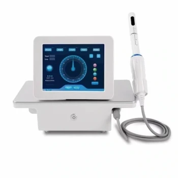
- Vaginal Tighten HIFU Gynecology HIFU Treatment
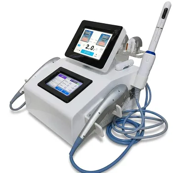
- Professional Face and Vaginal 7D HIFU System for HIFU Clinic Treatments
People Also Ask
- How does laser fluence influence pigment clearance vs. safety? Balancing Speed and Skin Integrity in Tattoo Removal
- What is the documented effectiveness of Q-switched Nd:YAG lasers for tattoo removal? Gold Standard Results
- What pigment compositions and particle properties should practitioners evaluate before operating Q-switched Nd:YAG lasers for tattoo removal? Evaluate pigment composition and particle size for safe, effective tattoo removal.
- How should aesthetic practitioners select wavelengths across Nd:YAG and Q-switched laser systems for target pigment depth and skin phototypes? Key Insights for Clinicians
- How should wavelength and fluence settings be adjusted on Nd:YAG and Q-switched lasers when treating darker skin tones? Optimize Safety & Efficacy