The primary benefit of using an 830 nm near-infrared diode laser for skin reflection imaging is its ability to penetrate deeply into tissue while causing minimal biological damage. This specific wavelength allows clinicians to visualize subsurface structures with high contrast, enabling the precise identification of pathologies that are invisible to the naked eye.
By leveraging the unique optical properties of skin at 830 nm, this technology turns natural melanin into a high-contrast beacon. This allows clinicians to visualize deep pathologies—such as Basal Cell Carcinoma and hyperpigmentation—without the need for invasive biopsies.
Optimizing Tissue Interaction
Achieving Deeper Penetration
Compared to shorter wavelengths in the visible spectrum, 830 nm light faces less scattering and absorption by water and hemoglobin.
This allows the laser to bypass surface barriers and probe deeper layers of the skin, providing a window into the dermis where many pathologies originate.
Minimizing Photodamage
Safety is a critical factor in diagnostic imaging. The 830 nm diode laser delivers the necessary optical power to generate clear images without causing significant photodamage to the delicate biological structures being observed.
Enhancing Diagnostic Accuracy
Detecting Basal Cell Carcinoma (BCC)
The primary clinical application of this technology is the detection of Basal Cell Carcinoma.
The laser generates sufficient backscattered signals to reveal tumor nests. When combined with acetic acid pretreatment, the system can clearly visualize BCC nests located in the deeper skin layers.
Evaluating Surgical Margins
Because the laser can delineate the boundaries of BCC nests, it is an invaluable tool for surgical planning.
Surgeons can perform a precise evaluation of surgical margins before making an incision, potentially reducing the need for repeat excisions and preserving healthy tissue.
Leveraging Melanin as a Natural Contrast Agent
At the 830 nm wavelength, melanin exhibits an extremely high refractive index.
This acts as a strong, natural endogenous contrast agent. Consequently, melanin-rich features appear with high brightness in the generated images, removing the need for external dyes in many pigment-focused assessments.
Applications in Pigmentary Analysis
Assessing Post-Inflammatory Hyperpigmentation (PIH)
The intense contrast provided by melanin allows for the quantitative assessment of pigmentary disorders.
Clinical systems can clearly identify the density of epidermal pigment and the brightness of the basal layer, offering objective data on the severity of Post-Inflammatory Hyperpigmentation.
Identifying Dermal Abnormalities
Beyond the surface, the high brightness of melanin at this wavelength highlights melanophages within the dermis.
This capability is essential for distinguishing between superficial epidermal pigmentation and deeper dermal pigment issues, which require different treatment approaches.
Understanding the Operational Trade-offs
Dependence on Contrast Enhancers for Oncology
While the laser is effective on its own for pigment analysis, detecting cancerous lesions like BCC requires a specific protocol.
The laser alone may not provide sufficient differentiation for tumor nests; acetic acid pretreatment is a requisite step to create the necessary contrast. Clinicians must adhere to this multi-step workflow to ensure diagnostic accuracy.
Making the Right Choice for Your Goal
The utility of an 830 nm laser source depends on the specific clinical pathology you are targeting.
- If your primary focus is Surgical Oncology (BCC): You will rely on the laser's deep penetration and acetic acid integration to map tumor nests and define precise surgical margins.
- If your primary focus is Dermatology (Pigmentation): You will utilize the high refractive index of melanin at this wavelength to quantify PIH and map dermal melanophages without chemical enhancers.
By selecting the 830 nm wavelength, you are prioritizing deep, non-destructive visualization to drive more accurate, evidence-based treatment decisions.
Summary Table:
| Feature | 830 nm NIR Laser Benefit |
|---|---|
| Tissue Penetration | Deep penetration into the dermis with minimal scattering |
| Biological Safety | Non-invasive imaging with extremely low risk of photodamage |
| Contrast Agent | Utilizes natural melanin as a high-refractive index beacon |
| Clinical Focus | Precision mapping of BCC nests and surgical margins |
| Pigment Analysis | Objective quantification of PIH and dermal melanophages |
Elevate Your Clinic with Advanced Diagnostic Precision
At BELIS, we specialize in professional-grade medical aesthetic equipment designed exclusively for high-end clinics and premium salons. Our advanced laser systems, including Diode Hair Removal, CO2 Fractional, Nd:YAG, and Pico lasers, leverage the latest optical technologies to deliver superior clinical outcomes.
Whether you are looking for powerful body sculpting solutions like EMSlim and Cryolipolysis or specialized care devices such as HIFU, Microneedle RF, and skin testers, our portfolio provides the reliability and precision your practice demands.
Ready to upgrade your diagnostic and treatment capabilities? Contact us today to explore our full range of professional systems
References
- Yogesh G. Patel, Milind Rajadhyaksha. Confocal reflectance mosaicing of basal cell carcinomas in Mohs surgical skin excisions. DOI: 10.1117/1.2750294
This article is also based on technical information from Belislaser Knowledge Base .
Related Products
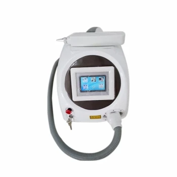
- Clinic Diode Laser Hair Removal Machine with SHR and Trilaser Technology
- Diode Laser SHR Trilaser Hair Removal Machine for Clinic Use
- 9D 7D HIFU Vaginal RF Lifting Treatment
- Vaginal Tighten HIFU Gynecology HIFU Treatment
- Trilaser Diode Hair Removal Machine for Beauty Clinic Use
People Also Ask
- What are the advantages of the Constant Motion Technique? Elevate Your Clinic's Laser Hair Removal Results
- Why can traditional high-energy pulse (HR) modes lead to adverse skin reactions? Understanding Thermal Injury Risks
- Why is 30 J/cm2 the threshold for diode laser hair removal? Ensure effective follicle destruction
- What is the necessity of trimming before Diode Laser hair removal? Essential Steps for Safety & Efficacy
- What is the primary mechanism of action for Diode Laser hair removal? Mastering Selective Photothermolysis