Facial anatomy determines both the result and the risk of HIFU or microneedle RF treatment. Practitioners must distinguish the epidermis, dermis, superficial and deep fat compartments, retaining ligaments, SMAS, muscles, bone, and major vessels before selecting treatment depth and energy. HIFU is designed to create focused thermal zones at selected tissue depths, while microneedle RF delivers heat through insulated or non-insulated needles into predetermined dermal or subdermal planes.
The key principle is anatomical precision: tightening should target the appropriate dermal or structural layer while preserving facial fat, vascular structures, nerves, and the epidermal barrier. Depths and energy settings must be individualized because facial tissue thickness varies substantially by region and patient.
Why Facial Tissue Layers Matter
The epidermis and dermo-epidermal junction
The epidermis is the superficial protective barrier and should not receive excessive thermal or mechanical injury. Beneath it, the dermo-epidermal junction contributes to adhesion between the epidermis and dermis through the basement membrane and hemidesmosomes.
Microneedle RF penetrates through the epidermis to deposit energy in the dermis, but incorrect insertion depth, excessive energy, or poor contact can still cause burns, scarring, pigmentary change, or barrier disruption.
The papillary and reticular dermis
The dermis contains a superficial papillary region and a deeper, structurally denser reticular region. The reticular dermis contains much of the collagen and elastin framework relevant to skin firmness.
For microneedle RF, the clinically important question is whether the selected needle depth places the thermal zone in the intended dermal layer without extending unnecessarily into fat. HIFU may also be configured to target deeper dermal planes, but the actual focal depth depends on the device and cartridge.
Superficial and deep facial fat
Facial subcutaneous fat is not a single uniform layer. It is organized into superficial and deep compartments separated by connective tissue planes, retaining ligaments, and the superficial musculoaponeurotic system.
The superficial fat compartments contribute to contour and skin mobility and may be more affected by body-weight changes. The deep fat compartments provide structural support, facilitate tissue gliding, and contribute to facial projection and volume.
The SMAS and deeper structures
The SMAS is a fibromuscular connective tissue layer that connects facial muscles with the superficial fascia and overlying soft tissue. It is clinically relevant to lifting procedures because force and contraction in this region can influence the overlying skin.
HIFU systems may offer focal depths intended to reach the deep dermis, subcutaneous tissue, or a fibromuscular plane such as the SMAS. However, practitioners should not assume that every device or cartridge reaches the SMAS accurately; treatment depth is device-specific and must be verified from the manufacturer’s technical documentation.
How Fat Distribution Changes Treatment Planning
Preserve necessary facial volume
Energy-based tightening can be inappropriate in naturally thin or volume-depleted areas. Excessive thermal exposure to superficial or deep fat may contribute to unwanted volume reduction, contour irregularity, or a more hollow appearance.
This is particularly important in the temples, periorbital region, anterior cheeks, and other areas where small volume changes can be visually prominent.
Aging changes more than skin laxity
Facial aging involves collagen loss, weakening of retaining ligaments, changes in fat volume and position, and reduction of underlying skeletal support. The apparent descent of tissue is therefore not corrected simply by treating the deepest available layer.
Treatment planning should identify whether the dominant issue is skin laxity, fat excess, fat deficiency, tissue descent, or loss of bony projection. A tightening device cannot reliably replace missing structural volume.
Regional thickness is highly variable
The cheek, malar region, jawline, temple, forehead, and periorbital areas have different skin thicknesses, fat compartments, vascular patterns, and tolerance for thermal injury. A setting that is appropriate for a thicker lower-face region may be excessive for a thin temple or bony area.
Palpation, visual assessment, and appropriate imaging or skin-analysis tools can help estimate regional tissue thickness and identify areas where a deep treatment would be unsuitable.
Matching the Device to the Anatomical Target
HIFU: focused treatment at selected depths
HIFU creates discrete thermal coagulation zones at a defined focal depth. Depending on the system, available depths may include superficial dermal targets and deeper subcutaneous or fascial targets.
The practitioner must confirm that the focal zone is positioned in the intended tissue plane. Deeper is not automatically better: placing energy too superficially may injure the skin, while placing it too deeply or repeatedly may affect fat, muscle, nerves, or other structures.
Microneedle RF: needle depth and thermal spread
Microneedle RF uses mechanical needle insertion followed by radiofrequency heating at or around the needle tips. The relevant anatomical variables are the insertion depth, electrode design, energy delivery pattern, pulse duration, and thermal spread beyond the needle tips.
A nominal needle depth does not always equal the final tissue-heating depth. Tissue compression, insertion angle, needle configuration, impedance, and energy settings can all alter the effective treatment zone.
Depth labels are not universal anatomical guarantees
Depths such as 1.5 mm, 3.0 mm, or 4.5 mm are commonly discussed in energy-based treatment, but they should not be treated as universal prescriptions. Device calibration, focal-zone geometry, patient anatomy, and manufacturer-specific terminology differ.
The correct approach is to select the shallowest effective target for the intended clinical goal and verify that the device’s treatment plane corresponds to the patient’s actual tissue anatomy.
High-Risk Anatomical Regions
Temple and lateral forehead
The temple contains branches of the superficial temporal artery and important fascial planes. The depth and course of vessels can vary, and the region may have limited soft-tissue thickness over bone.
Deep energy delivery or needle insertion in this area therefore requires conservative planning, accurate anatomical knowledge, and strict adherence to device-specific safety guidance.
Glabella and medial forehead
The glabellar region contains interconnected branches of the supratrochlear, supraorbital, and dorsal nasal arterial systems. Thermal or mechanical injury in this region carries greater potential consequence than routine treatment in a thicker, less vascular area.
Practitioners should avoid assuming that a superficial appearance indicates a simple anatomy. The location of vessels and the risk of unintended injury must be considered before selecting depth, energy, and treatment pattern.
Periorbital and other thin zones
The periorbital region has thin soft tissue and important underlying structures. Aggressive treatment can produce pain, swelling, contour changes, or injury to structures that are close to the treatment field.
Settings and treatment indications should be based on the specific device’s approved use and the clinician’s training, rather than extrapolated from cheek or jawline protocols.
Areas near nerves and muscle
Facial motor nerve branches travel through variable tissue planes, particularly around the temple, cheek, lower face, and jawline. Although HIFU and RF are intended to act within selected tissue layers, inaccurate depth or excessive thermal exposure may increase the risk of transient or persistent nerve-related effects.
Anatomical mapping should therefore include not only fat and fascia, but also the expected course of nerves and muscles.
Patient-Specific Factors That Alter Depth Selection
Thin versus thick soft tissue
A thin face offers less separation between the skin, fat, muscle, fascia, bone, vessels, and nerves. The same nominal treatment depth can therefore have very different consequences in two patients.
Thickness should be assessed region by region rather than inferred from overall body size or facial appearance alone.
Sex, age, and hormonal changes
Average differences in skin thickness and collagen content exist between individuals, but sex-based assumptions are not sufficient for setting treatment parameters. Age, hormonal status, prior procedures, sun exposure, medications, and disease can be equally or more important.
Each patient should be assessed individually rather than assigned a deeper or shallower protocol solely because of sex.
Previous procedures and scarring
Fillers, threads, surgery, implants, fat grafting, inflammation, and scar tissue can alter tissue planes and mechanical resistance. Scarred or poorly vascularized areas may respond differently and may have less predictable healing.
A complete history and examination are essential before treating areas that have undergone previous intervention.
Understanding the Trade-offs
Tightening versus volume preservation
The desired result is usually improved firmness and contour without loss of healthy facial volume. Treating too broadly, too deeply, or too frequently can shift the balance toward hollowing or contour irregularity.
This is why deep fat should not be treated merely because it is accessible. The intended tissue target must be linked to a specific clinical problem.
More energy does not guarantee more lifting
Increasing energy or stacking passes may increase thermal injury without producing proportionally greater tightening. Collagen remodeling is biologically limited, and excessive treatment can cause inflammation, pain, burns, scarring, pigmentary changes, or fat alteration.
Safe treatment requires controlled thermal exposure, adequate spacing, and respect for regional anatomy.
Anatomic landmarks are helpful but imperfect
Surface landmarks provide a starting point, not a complete map. Vascular, neural, fascial, and fat-compartment anatomy varies between patients and can be altered by aging or prior treatment.
When the treatment area is thin, previously treated, asymmetric, or high-risk, additional assessment may be more valuable than simply changing device settings.
HIFU and microneedle RF are not interchangeable
HIFU creates focused energy zones at selected depths, whereas microneedle RF creates heat around inserted electrodes. Their tissue effects, depth control, thermal distribution, and risk profiles are different.
A protocol designed for one technology should not be transferred directly to the other based only on a similar numerical depth.
How to Apply This to Clinical Planning
Anatomical assessment should precede device selection, depth selection, and energy adjustment.
- If your primary focus is skin texture and superficial dermal remodeling: Favor a controlled dermal target with microneedle RF or an appropriate superficial HIFU setting, while protecting the epidermis and avoiding unnecessary extension into fat.
- If your primary focus is laxity and structural support: Assess the reticular dermis, subcutaneous compartments, retaining ligaments, and SMAS before selecting a deeper focal plane; use only device-validated depths.
- If your primary focus is preserving facial volume: Identify thin or hollow areas and avoid indiscriminate treatment of superficial or deep fat, particularly where small volume changes would be conspicuous.
- If your primary focus is treating the temple, glabella, or periorbital region: Treat these as high-anatomical-risk zones and use conservative, anatomy-based planning by an appropriately trained clinician.
- If your primary focus is individualized treatment safety: Document regional tissue thickness, prior procedures, asymmetry, vascular and neural risk, and the intended tissue target before treatment.
The safest and most effective energy-based facial treatment is guided by the tissue plane that needs improvement—not simply by the deepest cartridge or highest available energy.
Summary Table:
| Anatomical Structure | Relevance to HIFU/Microneedle RF | Key Considerations |
|---|---|---|
| Epidermis & Dermo-epidermal Junction | Must be protected from thermal injury | Risk of burns, scarring, pigmentary changes if damaged |
| Papillary & Reticular Dermis | Main targets for tightening | Depth selection determines collagen remodeling; avoid excessive energy |
| Superficial & Deep Fat Compartments | Preserve volume | Risk of fat loss and contour irregularities if targeted indiscriminately |
| SMAS & Deeper Structures | May be targeted for lifting | Depth must be device-specific; not all devices reach SMAS |
| High-Risk Regions (Temple, Glabella, Periorbital) | Require conservative planning | Vessels, nerves, thin tissue; higher risk of adverse events |
| Patient-Specific Factors (Thickness, Age, Prior Procedures) | Individualize treatment parameters | Avoid assumptions; assess each patient individually |
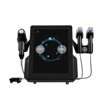
Elevate your practice with BELIS's advanced HIFU and microneedle RF systems, designed for precision and safety. Our devices offer customizable depth settings and rigorous safety features to help you achieve optimal outcomes for your patients. Whether you're a clinic or premium salon, our dedicated support and OEM/ODM options ensure a seamless integration into your services. Ready to enhance your aesthetic offerings? Contact us today to learn how BELIS can empower your medical spa and delight your clients with cutting-edge technology and exceptional results.
Related Products
- RF Microneedling Machine Micro Needle Radio Frequency Machine
- RF Microneedling Machine Micro Needle Radio Frequency Machine
- 9D 7D HIFU Vaginal RF Lifting Treatment
- 4D Vaginal HIFU and Face HIFU System
- Professional Face and Vaginal 7D HIFU System for HIFU Clinic Treatments
People Also Ask
- How does Microneedling assist in drug absorption for vitiligo? Boost Treatment Efficacy with Enhanced Micro-channels
- How does Microneedling RF differ from standard microneedling? Advanced Scars Treatment for Clinic Results
- What is the function of applying high-concentration topical anesthetic cream? Unlock Deeper Results in Microneedling
- Why is evaluating localized skin thickness essential when setting needle depth and energy parameters for microneedling RF and microdermabrasion devices? Achieve Safe and Effective Treatments
- Why is a 2.5mm needle depth typically selected for deep scars? Unlock Professional Microneedling Results