Clinics should manage post-laser erythema by distinguishing expected healing from a complication, then tailor treatment to the patient’s skin type, procedure, and wound status. After ablative resurfacing, erythema may persist for one to four months as blood flow and tissue regeneration remain elevated. Redness that is worsening, intensely pruritic, painful, blistering, malodorous, or accompanied by drainage or fever requires prompt evaluation for contact dermatitis, infection, or excessive thermal injury.
The key is trajectory and timing: stable, gradually improving erythema can be part of normal recovery, while intensifying or unusually persistent inflammation should trigger a clinical assessment. Pigmentary complications are managed primarily through complete wound healing, strict photoprotection, supervised pigment-modulating therapy, and conservative laser technique.
Establish Whether the Reaction Is Expected
Assess the procedure and treatment depth
The expected course depends on whether treatment was ablative or non-ablative, the device and settings, the number of passes, pulse stacking, treatment density, and the patient’s baseline vascularity and skin type.
Ablative resurfacing commonly produces more prolonged erythema than non-ablative treatment. The clinic should document these expectations before treatment so that patients understand that improvement may take months rather than days.
Focus on the direction of change
A gradual reduction in redness is generally reassuring. Increasing erythema, new pruritus, worsening pain, blistering, erosions, or purulent drainage is not simply “normal redness” and should be assessed.
Persistent erythema is more concerning when it appears disproportionate to the treatment, follows exposure to a new product, or develops alongside systemic symptoms.
Look for common causes of excessive redness
The principal causes to consider include:
- Contact dermatitis from fragrances, botanical extracts, harsh additives, or non-prescribed skincare products.
- Bacterial, fungal, or viral infection, including herpes simplex reactivation.
- Excessive thermal injury from high density, long pulse widths, multiple passes, or pulse stacking.
- Vascular reactivity, particularly in patients with telangiectasia or pre-existing redness.
- Occlusive or otherwise inappropriate aftercare that increases irritation or microbial risk.
Manage Erythema Without Delaying Healing
Remove potential irritants first
If contact dermatitis is suspected, instruct the patient to stop the suspected product immediately and return to the clinic’s prescribed, minimal aftercare regimen.
The recovery routine should be fragrance-free, hypoallergenic, and limited to products appropriate for the current wound-healing stage. Patients should not add botanicals, exfoliants, retinoids, bleaching products, or active ingredients without clinical approval.
Examine for infection before suppressing inflammation
Before prescribing anti-inflammatory treatment for severe or worsening erythema, clinicians should evaluate for active bacterial, fungal, or viral infection.
Unexplained pain, warmth, spreading redness, drainage, fever, vesicles, or rapidly worsening symptoms warrant prompt medical review. Antiviral prophylaxis may be appropriate for patients at risk of recurrent herpes simplex, according to the treating clinician’s protocol.
Use corticosteroids selectively
A clinician may consider a low-potency topical corticosteroid or, in selected severe cases, a short systemic corticosteroid course when excessive inflammation or hypersensitivity is diagnosed and infection has been excluded.
However, corticosteroids should not be used as a routine substitute for diagnosis. Application to incompletely healed or re-epithelialized skin requires particular caution because inappropriate use can delay closure or increase infection risk.
Protect the recovering skin
Patients should avoid direct sun exposure, heat, saunas, strenuous exertion, alcohol, and spicy foods when these worsen flushing or discomfort.
Once the skin has fully re-epithelialized, broad-spectrum UVA/UVB protection becomes essential. Pure mineral formulations containing zinc oxide or titanium dioxide may be useful because they provide physical protection and are generally well tolerated when applied appropriately.
Add camouflage only after re-epithelialization
Camouflage makeup should not be applied over open, eroded, or incompletely healed skin. After the barrier is restored, fragrance-free mineral makeup can reduce the visible burden of erythema and improve patient confidence.
Green- or yellow-toned correctors can visually neutralize red undertones, followed by a light mineral base. This is a cosmetic adjunct, not treatment for the underlying inflammation.
Prevent and Treat Post-Inflammatory Hyperpigmentation
Identify patients at higher risk
Post-inflammatory hyperpigmentation is particularly common in darker Fitzpatrick skin types and can be intensified by prolonged inflammation and ultraviolet exposure.
Patients with darker phototypes, a history of hyperpigmentation, recent tanning, or significant sun exposure require especially careful counseling and conservative treatment planning.
Make photoprotection non-negotiable
Daily, broad-spectrum UVA/UVB sunscreen and strict avoidance of direct solar exposure are central to pigment management. Clinics should provide written instructions and explain that even modest ultraviolet exposure during active inflammation can intensify pigmentation.
Photoprotection should continue after visible erythema improves because pigmentary activity may persist after the surface appears healed.
Start pigment-modulating therapy only on healed skin
After complete re-epithelialization, clinicians may consider topical hydroquinone and, when appropriate, a topical retinoid to manage post-inflammatory hyperpigmentation.
These agents can irritate recently treated skin, so they should be introduced only when the barrier is adequately restored and under a clinician-directed schedule. Irritation itself can worsen pigmentation, making gradual use and close follow-up important.
Treat persistent vascular redness separately
Persistent redness and hyperpigmentation are related but not identical problems. If vascular erythema remains after the active healing phase, targeted vascular treatments such as intense pulsed light or pulsed dye laser therapy may be considered.
Treatment timing and parameters must account for the patient’s phototype, residual sensitivity, and risk of further pigment alteration. These modalities should not be used reflexively while the skin remains unstable.
Reduce the Risk of Hypopigmentation and Scarring
Prevention begins during treatment
Delayed hypopigmentation can follow deep ablation or removal of severely sun-damaged tissue. The risk increases with overly aggressive settings, excessive passes, and inadequate control around delicate areas such as the perioral region.
Meticulous peripheral feathering and conservative energy delivery help reduce abrupt treatment borders and excessive thermal injury.
Set realistic expectations for treatment
Post-treatment hypopigmentation can be difficult to correct and may not respond predictably to intervention. Specialized devices, including 308-nm excimer systems, may be considered in selected cases by clinicians experienced in treating this complication.
The decision should follow an assessment of whether pigment loss is stable, whether scarring is present, and whether further treatment could create additional contrast or inflammation.
Understanding the Trade-offs
Do not treat every red face aggressively
Prolonged erythema can be a normal component of ablative healing. Premature use of additional lasers, potent actives, or aggressive anti-inflammatory therapy may disrupt healing or create new complications.
The safer approach is to monitor the trend, exclude complications, and intervene according to the cause.
Do not assume all persistent redness is infection
Infection must be ruled out when clinically indicated, but unnecessary antimicrobial or antibiotic use can cause contact dermatitis and further confuse the presentation.
The clinic should base treatment on examination and, where appropriate, diagnostic testing rather than treating redness alone.
Balance inflammation control against wound repair
Inflammation is part of tissue repair, but excessive inflammation can prolong erythema and promote hyperpigmentation. Anti-inflammatory medication may be appropriate in selected cases, yet its timing and strength must be balanced against barrier recovery and infection risk.
Avoid unvalidated or poorly timed add-ons
Supportive measures such as microcurrent therapy, vascular gels, or additional light-based procedures should not replace diagnosis, photoprotection, and appropriate wound care. Evidence, timing, device parameters, and patient selection should be considered before adding them to a recovery protocol.
Build compliance into the clinical process
Patients often worsen pigmentary outcomes through sun exposure or unsupervised product use rather than through the procedure alone. Clinics should provide clear written instructions, obtain documented consent, supply contact information for concerns, and use standardized photographs to monitor progress.
How Clinics Should Apply This Protocol
The most reliable approach is a staged pathway based on healing status and clinical findings:
- If your primary focus is expected erythema: Explain that ablative resurfacing may produce redness for one to four months, while requiring review if symptoms intensify or fail to improve progressively.
- If your primary focus is severe or worsening erythema: Stop non-prescribed products, evaluate for dermatitis, infection, and thermal injury, and prescribe anti-inflammatory treatment only after the diagnosis and wound status are established.
- If your primary focus is post-inflammatory hyperpigmentation: Begin strict UVA/UVB protection immediately as appropriate for the healed surface, then consider supervised hydroquinone and topical retinoid therapy after re-epithelialization.
- If your primary focus is persistent vascular redness: Allow adequate recovery, then consider targeted vascular laser or IPL treatment using parameters appropriate for the patient’s phototype and sensitivity.
- If your primary focus is hypopigmentation prevention: Use conservative energy settings, avoid excessive passes, and feather treatment margins carefully, particularly in delicate or heavily sun-damaged areas.
- If your primary focus is patient adherence: Give simple written aftercare instructions, prohibit unsupervised active products, document counseling, and arrange follow-up before the patient leaves the clinic.
With disciplined assessment, conservative wound care, and strict photoprotection, clinics can distinguish normal recovery from complications and manage pigmentary changes without creating additional injury.
Summary Table:
| Aspect | Key Management Points |
|---|---|
| Erythema Duration | Normal after ablative resurfacing: 1-4 months; gradual improvement expected. |
| Red Flags | Worsening redness, pruritus, pain, blistering, drainage, fever → assess for dermatitis, infection, thermal injury. |
| Contact Dermatitis | Stop suspected products; simplify aftercare to fragrance-free, hypoallergenic regimen. |
| Infection | Rule out bacterial, fungal, viral (herpes) before anti-inflammatory treatment; consider antiviral prophylaxis. |
| Corticosteroids | Use low-potency or short systemic course only after infection excluded; cautious on healing skin. |
| Photoprotection | Strict UVA/UVB protection (mineral sunscreen) after re-epithelialization; crucial for PIH prevention. |
| Camouflage | Only after re-epithelialization; use green/yellow correctors and mineral makeup. |
| Post-Inflammatory Hyperpigmentation | Identify high-risk patients (darker skin types); initiate hydroquinone/retinoid only on healed skin; avoid irritation. |
| Vascular Redness | After healing, consider IPL or pulsed dye laser for persistent erythema. |
| Hypopigmentation | Prevent with conservative laser settings, peripheral feathering; treat with 308-nm excimer if stable. |
| Patient Education | Provide written instructions, obtain consent, use photos, and follow up for compliance. |
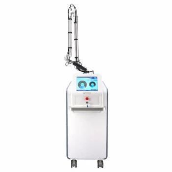
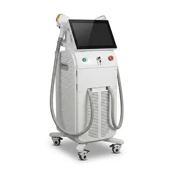
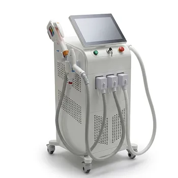
Elevate your clinic's post-laser care with BELIS's advanced aesthetic technology. Our professional-grade laser systems (Diode, Alexandrite, CO2, Nd:YAG) and complementary devices ensure precise, safe treatments that minimize side effects. Contact us today at #ContactForm to learn how BELIS can enhance your practice's outcomes and patient satisfaction.
Related Products
- Fractional CO2 Laser Machine for Skin Treatment
- Fractional CO2 Laser Machine for Skin Treatment
- Pico Laser Tattoo Removal Machine Picosure Picosecond Laser Machine
- Clinic Use IPL SHR ND YAG Laser Hair Removal RF Skin Tightening Machine
- Diode Tri Laser Hair Removal Machine for Clinic Use
People Also Ask
- What is the technical principle behind CO2 Laser Fractional micro-perforations? Master Scar Revision Mechanics
- Why are professional laser systems, such as CO2 fractional or Erbium lasers, more effective for deep wrinkle reduction and dermal remodeling than topical anti-aging ingredients like retinoids, vitamin C, or peptides? Discover the targeted power of fractional lasers.
- What role does fractional CO2 laser equipment play in the treatment of SUI? Non-Surgical Stress Urinary Incontinence Care
- What is the rationale for a double-pass technique with fractional CO2 lasers? Maximize Deep Collagen Remodeling
- What is the purpose of manually extracting large cysts before CO2 fractional laser? Optimize Eyelid Milia En Plaque Care