High-BMI patients undergoing body contouring require a formal airway and sedation-risk assessment before treatment begins. Clinicians should assess facial anatomy, mouth opening, neck mobility, thyromental distance, likely obstructive sleep apnea, aspiration risk, and the planned depth and duration of anesthesia. Findings that suggest a difficult airway should trigger early anesthesia involvement, appropriate equipment, a rescue plan, and consideration of a setting capable of advanced airway management.
The central principle is preparation, not prediction. Airway examination findings are warning signals rather than guarantees; high-BMI patients should be managed with a documented primary airway plan, backup oxygenation and ventilation strategies, trained personnel, and clear escalation or transfer criteria.
Why Body Contouring Creates Additional Airway Risk
High BMI is a risk marker, not a complete airway diagnosis
Higher BMI is associated with reduced respiratory reserve, difficult mask ventilation, obstructive sleep apnea, and more rapid oxygen desaturation during apnea. These risks may be amplified by prolonged procedures, supine positioning, sedatives, opioids, abdominal compression, and postoperative swelling.
BMI alone cannot determine whether intubation or ventilation will be difficult. It should prompt a more comprehensive assessment rather than serve as the only screening criterion.
Procedure scale changes the safety requirements
Large-volume body contouring can involve prolonged anesthesia, substantial fluid administration, temperature loss, and postoperative third-space fluid shifts. These factors increase the importance of controlled monitoring and a reliable plan for airway support after the procedure.
For large-volume procedures, particularly when more than 5 liters are aspirated, the clinic should follow applicable surgical and anesthesia regulations regarding observation, fluid management, warming, and postoperative monitoring. A fixed observation period should not replace individualized clinical assessment.
How to Screen for a Potentially Difficult Airway
Inspect mouth opening and oral anatomy
Measure the interincisor distance. An opening of less than approximately 3 cm is concerning because it may restrict laryngoscopy and placement of airway devices.
Also assess:
- Mallampati class III or IV
- An inability to visualize the uvula
- Prominent upper incisors
- A substantial overbite
- Limited temporomandibular-joint movement
- Facial hair or anatomy that may interfere with a mask seal
No single finding reliably predicts difficulty. The concern increases when several abnormalities occur together.
Assess the neck and jaw
Evaluate neck circumference, neck length, cervical flexion and extension, and the ability to position the head and neck appropriately.
A very large neck circumference—particularly measurements around 60 cm—is a significant warning sign. Thresholds should be interpreted in context rather than applied as universal sex-specific cutoffs, because neck size, body habitus, and airway anatomy vary substantially.
Assess the thyromental distance as well. A distance of less than approximately three fingerbreadths may indicate limited mandibular space and a more difficult laryngoscopic view.
Screen for obstructive sleep apnea
Ask about loud snoring, witnessed apneas, nocturnal choking, morning headaches, daytime somnolence, and previous use of continuous positive airway pressure. Use a validated screening approach, such as STOP-Bang, where appropriate.
A known or suspected sleep-related breathing disorder should influence the sedation plan, postoperative observation, opioid use, and decision about whether the clinic is an appropriate location for the procedure.
Review previous airway and anesthesia history
Previous difficult intubation, difficult mask ventilation, unexpected intensive-care admission, or postoperative airway obstruction is highly relevant. Obtain prior anesthesia records when possible rather than relying only on the patient’s recollection.
Also identify aspiration risks, such as reflux, delayed gastric emptying, diabetes with gastroparesis, opioid use, and inadequate fasting.
How to Build the Airway Plan Before Sedation
Match the procedure to the facility
A clinic should only provide the level of anesthesia that its personnel, equipment, monitoring, recovery area, and emergency-transfer arrangements can safely support.
Patients with multiple difficult-airway predictors, severe or untreated obstructive sleep apnea, significant cardiopulmonary disease, or anticipated deep sedation may require an anesthesiologist-led plan or a hospital or accredited ambulatory surgical facility.
Define primary and rescue strategies
The team should document:
- The intended level of anesthesia and ventilation.
- The primary airway technique.
- The backup method for oxygenation and ventilation.
- The plan for failed intubation or failed ventilation.
- Who will call for additional help.
- The criteria for aborting the procedure or transferring the patient.
A difficult-airway cart should be immediately accessible and checked before each case. It should include appropriately sized face masks, oral and nasal airways, supraglottic airways, video laryngoscopy equipment, fiber-optic equipment when indicated, suction, capnography, and equipment for emergency front-of-neck access according to local standards.
Optimize positioning and preoxygenation
Positioning a high-BMI patient with the upper body elevated and the head and neck aligned can improve preoxygenation, mask ventilation, and laryngoscopy. The exact position should be individualized and maintained during induction when appropriate.
Preoxygenation should be deliberate and confirmed with appropriate monitoring. Because these patients may desaturate quickly, the team should minimize unnecessary apnea and ensure that rescue oxygenation is immediately available.
Managing Sedation and Airway Obstruction
Do not allow “moderate sedation” to become unintended deep sedation
Sedatives and opioids can convert partial upper-airway obstruction into apnea, especially in patients with obstructive sleep apnea. Titration should be conservative, with continuous observation and appropriate monitoring of ventilation, not merely oxygen saturation.
Capnography is particularly valuable because oxygen supplementation can delay recognition of hypoventilation or apnea when clinicians rely on pulse oximetry alone.
Use simple airway maneuvers early
For mild obstruction during sedation, perform a jaw thrust or gentle anterior mandibular advancement, reposition the head and neck, provide supplemental oxygen, and use oral or nasal airway adjuncts when appropriate and tolerated.
Do not wait for profound desaturation. If obstruction persists, deepen the airway intervention, assist ventilation, stop sedative administration, and escalate according to the predefined rescue plan.
Preserve spontaneous ventilation when a difficult airway is anticipated
In selected anticipated difficult-airway cases, maintaining spontaneous ventilation during airway establishment can provide an important margin of safety. However, this is not an automatic rule: spontaneous breathing may be inadequate, and coughing, movement, or obstruction may make the procedure unsafe.
The technique should be selected and performed by clinicians experienced in advanced airway management. Neuromuscular blockade should not be administered reflexively before the team has confirmed that it can oxygenate and ventilate the patient and has a credible rescue strategy.
Treat supraglottic airways as rescue or primary devices only when appropriate
Multiple sizes of laryngeal mask and other supraglottic airway devices should be available. They can provide effective ventilation in many circumstances, but they do not eliminate aspiration risk and may be less reliable in certain body positions, with high airway pressures, or during procedures that limit access to the airway.
Their role should be determined by the anesthetic plan, procedure characteristics, aspiration risk, and the operator’s expertise.
Postoperative Airway Management Matters
Recovery is a high-risk period
Airway obstruction and hypoventilation may occur after the procedure as sedatives and analgesics continue to act. Patients with suspected or known obstructive sleep apnea require careful observation until they are awake, maintaining oxygenation and ventilation, and able to protect their airway.
Use the patient’s prescribed positive-airway-pressure device when clinically appropriate. Opioid-sparing analgesia is preferable when feasible.
Plan observation according to risk
Patients undergoing extensive procedures, receiving substantial sedatives or opioids, or experiencing airway events should not be discharged solely because they are awake initially. The team should consider procedure magnitude, comorbidities, oxygen requirement, respiratory events, pain control, and the availability of responsible adult supervision.
For large-volume procedures, overnight or extended monitoring may be appropriate, but the duration should follow the patient’s condition and applicable regulations rather than an arbitrary time alone.
Understanding the Trade-offs
Airway testing has limited predictive power
Mallampati class, mouth opening, neck circumference, and thyromental distance can identify risk but cannot reliably exclude a difficult airway. A patient with a reassuring examination may still present unexpected difficulty.
Conversely, an abnormal finding does not automatically require cancellation. It requires an appropriately skilled team, equipment, environment, and escalation plan.
Spontaneous ventilation is not universally safer
Preserving spontaneous breathing can be valuable in an anticipated difficult airway, but it may also provide inadequate ventilation or poor operating conditions. The safest approach depends on the patient, procedure, airway technique, and clinician expertise.
Supraglottic devices do not replace a definitive airway plan
An LMA can rescue ventilation, but it is not equivalent to a secured tracheal airway and may not protect against aspiration. Clinics should not rely on an LMA as their sole difficult-airway strategy.
High-volume contouring requires broader physiologic planning
Airway safety cannot be separated from temperature control, fluid administration, local-anesthetic dosing, positioning, and postoperative observation. Tumescent lidocaine and epinephrine doses should be calculated from the patient’s characteristics, medical history, total planned dose, and institutional protocols.
How to Apply This to Your Clinic
Use a written checklist and require an airway briefing before every higher-risk body-contouring case.
- If your primary focus is preoperative safety: Document mouth opening, Mallampati class, neck anatomy and mobility, thyromental distance, temporomandibular movement, sleep-apnea risk, aspiration risk, and previous anesthesia problems.
- If your primary focus is procedural-site selection: Refer patients with multiple difficult-airway predictors, severe sleep apnea, major comorbidity, or anticipated deep sedation to an anesthesia-led environment with advanced rescue capability.
- If your primary focus is emergency readiness: Keep a checked difficult-airway cart immediately available, including video or fiber-optic equipment, supraglottic airways, suction, capnography, and a documented escalation plan.
- If your primary focus is sedation management: Titrate sedatives conservatively, monitor ventilation continuously, preserve spontaneous ventilation when clinically appropriate, and intervene early at the first sign of obstruction.
- If your primary focus is postoperative safety: Observe patients until respiratory function is stable and discharge criteria are met, with heightened vigilance after extensive procedures, opioid use, or airway events.
A safe high-BMI body-contouring program treats airway assessment, rescue preparation, and postoperative observation as one continuous process—not as separate tasks.
Summary Table:
| Risk Factor | Assessment | Red Flags |
|---|---|---|
| Mouth opening | Interincisor distance | < 3 cm |
| Oral anatomy | Mallampati class, visualization of uvula | Class III/IV, inability to see uvula |
| Neck & jaw | Neck circumference, thyromental distance | Neck circumference ~60 cm, thyromental distance < 3 fingerbreadths |
| OSA risk | STOP-Bang questionnaire | High score (≥3) or known OSA |
| Previous airway issues | Anesthesia records | History of difficult intubation or ventilation |
At BELIS, we prioritize patient safety. Our advanced body contouring devices, including laser systems and body sculpting technologies, are designed for use in professional settings. Ensure your clinic is equipped with the knowledge and tools to manage high-BMI patients safely. Contact us today to learn more about our state-of-the-art equipment and how we can support your practice.
Related Products
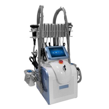
- Fat Freezing Cryolipolysis Machine for Body Contouring
- Fat Freezing Cryolipolysis Machine for Body Contouring
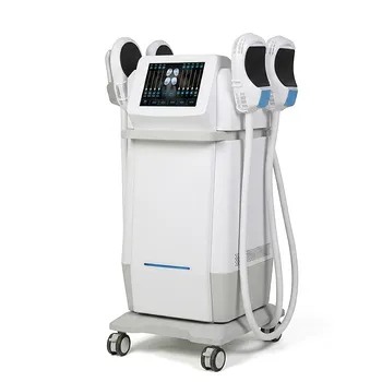
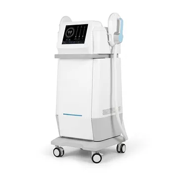
- EMSlim Neo Nova Body Sculpting EMS Sculpting Machine
- EMS Body Sculpting Machine for Body Slimming and Sculpting
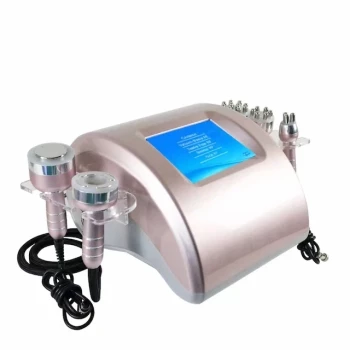
- Cryolipolysis Fat Freezing Machine and Ultrasonic Cavitation Device
People Also Ask
- What are the main benefits of using a fat freezing machine? Achieve Permanent, Non-Surgical Body Sculpting
- Do fat freezing machines really work? Achieve Non-Invasive Body Contouring
- What are the potential side effects and risks associated with fat freezing? Safety Profile & Rare Risks Explained
- What are the clinical and safety risks of using uncertified or replicate cryolipolysis body contouring equipment? Avoid hyperpigmentation and legal exposure with certified systems.
- What maintenance and service aspects are important when purchasing a fat freezing machine? Maximize ROI & Reliability