A multi-layer numerical skin model serves as a high-fidelity digital twin that quantifies the interaction between laser energy and complex biological structures. By simulating light propagation and thermal diffusion across distinct layers—such as the epidermis, dermis, and subcutaneous fat—it identifies critical safety thresholds and potential tissue damage risks before a device ever touches human skin.
A multi-layer numerical skin model provides a repeatable, risk-free environment to establish safety boundaries for aesthetic lasers. By calculating energy distribution across varied tissue depths, it allows manufacturers to validate equipment safety under extreme parameters without the immediate need for clinical trials.
Precision Simulation of Tissue Interaction
Mapping Structural and Physical Parameters
The model functions by accurately defining the structural characteristics of the epidermis, dermis, and subcutaneous fat. This includes the integration of various vascular networks to mimic real-world biological complexity.
By assigning specific physical properties to these layers, the model creates a predictable environment for testing. This level of detail is essential for understanding how different tissues react to specific laser wavelengths.
Quantifying Light Propagation and Thermal Diffusion
The core utility of the model lies in its ability to perform quantitative calculations of light propagation paths. It tracks how photons move through tissue and where energy is ultimately absorbed.
Simultaneously, it simulates thermal diffusion, predicting how heat spreads from the target area to surrounding healthy tissue. This prevents "hot spots" that could lead to unintended burns or scarring.
Reducing Clinical Risk via Digital Pre-testing
Testing Extreme Operating Parameters
One of the most significant contributions to safety is the ability to simulate extreme operating parameters. Developers can push the digital model to its breaking point to see where tissue failure occurs.
This process establishes a "safety buffer" for the equipment. It ensures that the maximum settings available to a practitioner remain well within the limits of biological tolerance.
Eliminating Preliminary Clinical Hazards
Because the model is a digital platform, it provides a highly controllable and repeatable environment. This reduces the ethical and physical risks associated with early-stage human testing.
By the time a device reaches clinical trials, its safety profile has already been rigorously "vetted" by the numerical model. This data-driven approach streamlines the path to regulatory approval while prioritizing patient safety.
Integrating Models with Real-World Diagnostics
Accounting for Melanin and Individual Variability
While the numerical model provides the framework, multi-functional diagnostic probes provide the specific data points needed for individual safety. These probes measure baseline melanin and erythema indices to account for varying skin tones.
Since melanin is the primary absorber of near-infrared light, these measurements are critical. The model can use this data to dynamically adjust laser dosage, compensating for high-absorption characteristics in darker skin types.
Real-Time Mapping and Guidance
Safety is further enhanced by real-time skin mapping systems that monitor the treatment area during the procedure. These systems guide the operator to adjust energy output in sensitive regions like the eye contour.
This integration prevents over-treatment in areas with heavy pigmentation and ensures sufficient energy in less sensitive zones. The result is a balanced treatment that minimizes the risk of post-operative complications.
Understanding the Trade-offs
Model Idealization vs. Biological Reality
Numerical models are, by definition, mathematical simplifications of highly complex biological systems. While they are incredibly accurate, they may not account for every idiosyncratic physiological response found in a diverse patient population.
Therefore, a model should never be the sole basis for safety. It must be used in conjunction with clinical observation and objective assessment tools like the Clinician Erythema Assessment (CEA).
Computational Intensity
High-fidelity multi-layer models require significant computational power to simulate complex thermal diffusion in real-time. There is often a trade-off between the depth of the simulation and the speed at which it can provide feedback to the laser operator.
How to Apply This to Your Safety Assessment
Implementing Model Data into Clinical Practice
- If your primary focus is Equipment Development: Use the multi-layer model to establish "hard" safety limits on energy density and pulse width before moving to human subjects.
- If your primary focus is Patient Safety: Rely on diagnostic probes to feed real-time melanin and erythema data into the pre-set models for personalized treatment calibration.
- If your primary focus is Treatment Efficacy: Utilize real-time skin mapping to ensure energy delivery is consistent and avoids both over-treatment and under-treatment.
By bridging the gap between digital simulation and clinical reality, multi-layer numerical models ensure that medical aesthetic lasers are both powerful enough to be effective and controlled enough to be safe.
Summary Table:
| Feature | Safety Contribution | Application |
|---|---|---|
| Structural Mapping | Identifies specific tissue reactions | Epidermis, dermis, and fat layer simulation |
| Thermal Diffusion | Prevents unintended burns and hot spots | Energy distribution and heat spread prediction |
| Digital Pre-testing | Establishes safety buffers | Testing extreme parameters without human risk |
| Diagnostic Integration | Personalizes energy dosage | Melanin and erythema index adjustments |
Elevate Your Clinic’s Safety with BELIS Advanced Technology
At BELIS, we specialize in professional-grade medical aesthetic equipment designed exclusively for clinics and premium salons that prioritize patient safety and clinical excellence. Our advanced laser systems—including Diode Hair Removal, Alexandrite, CO2 Fractional, Erbium, Nd:YAG, and Pico lasers—are engineered to deliver precise results while minimizing tissue risk.
From our HIFU and Microneedle RF skin tightening solutions to our powerful body sculpting portfolio (EMSlim, Cryolipolysis, RF Cavitation), we provide the digital precision and reliability your business needs to thrive.
Ready to upgrade your practice with industry-leading technology?
Contact BELIS Today to Request a Quote
References
- Yu Shimojo, Kunio Awazu. Picosecond laser-induced photothermal skin damage evaluation by computational clinical trial. DOI: 10.5978/islsm.20-or-08
This article is also based on technical information from Belislaser Knowledge Base .
Related Products
- Cryolipolysis Fat Freezing Machine and Ultrasonic Cavitation Device
- EMSlim RG Laser Body Sculpting and Slimming Machine
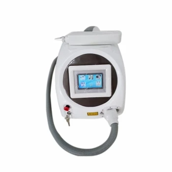
- Fractional CO2 Laser Machine for Skin Treatment
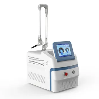
- Pico Picosecond Laser Machine for Tattoo Removal Pico Laser
- Pico Laser Tattoo Removal Machine Picosure Picosecond Laser Machine
People Also Ask
- Does cavitation get rid of belly fat? Yes, Here's How It Works for Body Sculpting
- Can ultrasonic waves destroy fat cells? A Guide to Non-Invasive Body Contouring
- How do Radio Frequency (RF) and Cryolipolysis devices achieve non-surgical body sculpting? Choose Your Best Tech
- How is a cryolipolysis procedure performed? A Step-by-Step Guide to Non-Invasive Fat Freezing
- How do Cryolipolysis devices achieve selective fat elimination? Unlock Non-Invasive Fat Cell Apoptosis Technology