Clinical facial lipoatrophy grading is based on how much contour volume has been lost, how prominent the underlying bones have become, and whether the facial musculature is visible. During consultation, practitioners assess affected regions such as the cheeks, temples, and periorbital areas, then assign a severity grade on a five-point clinical scale. The examination is supported by standardized photography, palpation, and, where available, skin diagnostic or 3D imaging systems to establish a treatment baseline.
Facial lipoatrophy is not graded by indentation alone. The most useful assessment combines contour flattening, bony landmark prominence, and visibility of underlying muscle, while also accounting for skin laxity, facial symmetry, and the patient’s treatment expectations.
How Practitioners Assess Facial Lipoatrophy
Evaluate the Face Under Standardized Conditions
The patient should be examined with the facial muscles relaxed and, where practical, in an upright or standing position. Consistent lighting, particularly directional lighting from above or the side, helps reveal shadows, depressions, and changes in facial topography.
Practitioners assess the face at rest and may also observe facial movement. A depression that appears only during muscle contraction may indicate dynamic dimpling or surface tethering rather than true generalized volume loss.
Examine Key Facial Regions
Assessment commonly includes the mid-cheeks, temples, periorbital hollows, preauricular region, and other areas of visible contour change. Each region should be considered separately because lipoatrophy may be localized, asymmetric, or more pronounced in one facial compartment than another.
The practitioner looks for flattening, concavity, shadowing, loose skin, changes in surface texture, and differences between the two sides of the face.
Use Palpation and Visual Comparison
Visual inspection identifies contour changes, while palpation helps estimate tissue thickness, firmness, mobility, and the relationship between the skin, subcutaneous tissue, and deeper structures. Comparison with the opposite side can help distinguish localized atrophy from normal anatomical asymmetry.
The consultation should also consider the patient’s age, prior weight changes, medical history, medications, previous procedures, and the timing of the perceived volume loss.
The Five-Grade Clinical Scale
Grade 1: Mild Flattening or Shadowing
Grade 1 represents mild volume loss. The practitioner may see subtle flattening or shadowing in areas such as the cheeks, temples, or periorbital region.
Bony landmarks are not prominently exposed, and the underlying musculature is not clearly visible. The change may be more apparent under directional lighting than during ordinary observation.
Grade 2: Early or Mild-to-Moderate Concavity
Grade 2 reflects a more defined contour depression than grade 1. The affected area may show early concavity and a clearer transition between the deficient region and adjacent tissue.
Bony prominence may begin to be noticeable, but the skeletal landmarks remain limited and muscle visibility is generally absent or minimal.
Grade 3: Moderate Concavity
Grade 3 indicates moderate facial lipoatrophy. The contour is visibly concave, and underlying bony structures become noticeable.
In some patients, the overlying tissue is sufficiently reduced that the outline or visibility of underlying musculature begins to appear. This grade commonly warrants detailed regional mapping before selecting a contouring approach.
Grade 4: Severe Volume Deficiency
Grade 4 represents advanced tissue loss approaching the most severe category. Indentation is pronounced, and bony landmarks are clearly prominent.
The skin may appear more closely draped over the underlying anatomy, with muscle visibility becoming more evident. Skin laxity and asymmetry should be documented because they may affect both treatment choice and the expected result.
Grade 5: Severe Indentation and Muscle Visibility
Grade 5 is the most severe category. It is characterized by extreme contour indentation, pronounced bony prominence, and clear visibility of the underlying musculature.
At this stage, the consultation must distinguish superficial appearance from substantial structural volume loss. Treatment planning should be conservative and anatomically precise, with realistic discussion of what a non-invasive procedure can and cannot achieve.
The Three Core Grading Criteria
Contour Flattening and Concavity
The first criterion is the degree to which the normal facial contour has flattened or become hollow. Practitioners assess both the depth of the depression and its length, width, borders, and relationship to neighboring facial compartments.
A broad, shallow hollow suggests a different structural problem from a small, sharply defined depression caused by focal tethering or scar-like fibrous bands.
Bony Landmark Prominence
As soft-tissue volume decreases, structures such as the orbital rim, zygomatic area, temporal ridge, or other skeletal landmarks may become more visible or palpable. Their prominence provides an important indication of the depth of soft-tissue deficiency.
Bony visibility should be interpreted in context because naturally lean facial anatomy can produce prominent landmarks without pathological lipoatrophy.
Visibility of Underlying Musculature
Visible muscle indicates a greater degree of tissue loss than surface shadowing alone. The practitioner should determine whether muscle visibility is present at rest, during expression, or under particular lighting conditions.
This distinction helps separate true structural volume deficiency from changes caused primarily by facial movement, lighting, or skin texture.
How Technology Supports the Clinical Examination
Standardized Photography
Standardized photographs provide a repeatable visual record of facial contours and asymmetry. Images should be taken with consistent lighting, facial expression, camera angle, and distance.
Photography is most useful when it complements a documented clinical examination rather than replacing direct assessment.
Skin Diagnostic Testing
Professional skin diagnostic equipment can help document surface characteristics, tissue condition, and visible concavities. These measurements may support baseline mapping before a treatment protocol is designed.
However, skin testing does not independently establish the clinical grade of lipoatrophy. The grade remains a clinical judgment based on anatomy, contour, and tissue volume.
Three-Dimensional Facial Imaging
Three-dimensional imaging can help quantify regional contour differences and document changes over time. It is particularly useful for identifying asymmetry and comparing pre-treatment and follow-up images under standardized conditions.
The technology should be treated as an adjunct because imaging accuracy depends on patient positioning, facial movement, lighting, software, and the quality of the acquisition process.
Distinguishing Lipoatrophy from Other Contour Problems
Diffuse Skin Laxity
Loose or poorly elastic skin can create sagging and shadowing that resembles volume loss. A practitioner should assess tissue turgor and elasticity separately from the amount of subcutaneous volume present.
Reducing tissue volume in an already lax area may worsen the appearance of deflation. The treatment plan may therefore need to address skin tightening rather than focus exclusively on fat reduction.
Focal Depressions and Fibrous Tethering
Small, clearly defined depressions may result from fibrous septa or localized tethering rather than generalized lipoatrophy. Their size, depth, borders, and behavior during movement should be documented.
Focal lesions of moderate severity may be candidates for procedures such as subcision when the clinical cause is appropriate. This is a different indication from diffuse facial volume deficiency.
Dynamic Dimpling and Surface Texture
A depression that appears only when a muscle contracts may reflect dynamic dimpling. Superficial “cottage cheese” texture and diffuse flaccidity may indicate a skin-surface or elasticity problem rather than substantial loss of deep facial volume.
These findings may favor approaches aimed at improving skin quality and elasticity, such as selected radiofrequency, HIFU, or laser protocols, when clinically appropriate.
Understanding the Trade-offs
Clinical Grading Is Structured but Not Entirely Mechanical
A five-grade scale improves consistency, but it does not eliminate professional judgment. Facial anatomy varies naturally, and the same visible hollow may have different causes in different patients.
Grades 1, 3, and 5 illustrate the mild, moderate, and severe endpoints described by the scale. Grades 2 and 4 represent intermediate findings and should be assigned according to how closely the patient’s presentation falls between those established levels.
Measurement Does Not Guarantee Treatment Suitability
A precise image or device reading can document a contour deficit, but it does not prove that a patient is suitable for a particular procedure. Safety, cause of the volume loss, tissue quality, medical history, and treatment goals remain central.
Technology is most valuable when it improves documentation and comparison without creating false certainty.
Expectations Must Be Assessed Alongside Anatomy
Patients may expect a procedure to restore youthful fullness, correct asymmetry, tighten loose skin, and improve texture simultaneously. Those goals may require different interventions and may not all be achievable with one modality.
Practitioners should explain the likely scope of improvement and decline treatment when expectations cannot be met realistically.
Insufficient Tissue Can Limit Contouring Options
When the area contains very little subcutaneous tissue, fat-reduction procedures may be inappropriate. Removing additional volume can intensify hollowness, skeletal prominence, or skin laxity.
The assessment should therefore confirm that the proposed procedure matches the patient’s actual tissue problem rather than the patient’s preferred treatment label.
Making the Right Choice for Your Goal
A sound consultation converts the grading result into an anatomically appropriate plan.
- If your primary focus is accurate severity grading: Document contour concavity, bony prominence, and muscle visibility for each affected region, using standardized examination conditions.
- If your primary focus is treatment planning: Determine whether the problem is diffuse volume loss, focal tethering, skin laxity, or dynamic dimpling before selecting a contouring modality.
- If your primary focus is progress evaluation: Combine standardized photographs, clinical examination, and consistent imaging or skin-testing methods to compare changes over time.
- If your primary focus is treatment safety: Review medical history, tissue quality, available subcutaneous volume, facial symmetry, and the realism of the patient’s expectations before proceeding.
A reliable lipoatrophy assessment links visible facial changes to their underlying anatomy, giving both practitioner and patient a defensible basis for treatment decisions.
Summary Table:
| Grade | Severity | Characteristics |
|---|---|---|
| 1 | Mild | Subtle flattening or shadowing; bony landmarks not prominent; muscle not visible. |
| 2 | Early | Definite concavity; early bony prominence; muscle visibility minimal. |
| 3 | Moderate | Visible concavity; bony structures noticeable; muscle may begin to show. |
| 4 | Severe | Pronounced indentation; clear bony prominence; muscle visibility evident. |
| 5 | Extreme | Severe indentation; pronounced bony landmarks; muscle clearly visible. |
Enhance your practice with advanced aesthetic devices from BELIS. Our portfolio includes cutting-edge laser systems (Diode, Alexandrite, CO2, Erbium, Nd:YAG, Pico), IPL, PDT, HIFU, Microneedle RF, body sculpting, Hydrafacial, and more—covering every category in aesthetic technology. Whether you are a clinic or premium salon, our professional-grade equipment supports precision and patient satisfaction. Contact our experts today to find the perfect solution for your contouring and rejuvenation needs. Contact us now for a personalized consultation and exclusive offers.
Related Products
- Hydrofacial Machine with Facial Skin Analyzer and Skin Tester
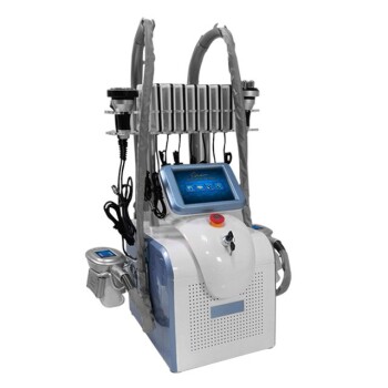
- Cryolipolysis Fat Freezing Machine Cavitation Lipo Laser Machine
- Fat Freezing Cryolipolysis Machine for Body Contouring
- Cryolipolysis Fat Freezing Machine with Cavitation and Laser Lipolysis
- Clinic Use IPL SHR ND YAG Laser Hair Removal RF Skin Tightening Machine
People Also Ask
- What is a HydroFacial treatment? Discover the Secret to Instant, Radiant Skin with Vortex-Fusion Technology
- What is the first step of a hydro-facial treatment? Master the Art of Professional Cleansing & Exfoliation
- What is a Hydrafacial machine and what are its unique features? Elevate Your Clinic with Vortex-Fusion Tech
- Is a HydraFacial machine worth it? Achieve Instant Glow with No Downtime
- What are the business benefits of a spa or salon owning a Hydrafacial machine? Drive Revenue and Loyalty