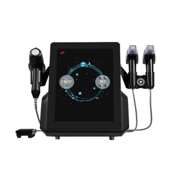
Use conservative, test-driven protocols. Nonablative fractional lasers treat striae by creating microscopic thermal treatment zones in the dermis, stimulating collagen remodeling while preserving the epidermis. Clinics should generally plan 2–4 sessions, use device-appropriate energy—commonly 35–55 mJ per microbeam/microbeam (mJ/MB) for standard tips—and reduce treatment coverage, often to approximately levels 4–7, when PIH risk is elevated. A test spot, strict photoprotection, and medically supervised pigment control are central to minimizing post-inflammatory hyperpigmentation.
The goal is controlled dermal remodeling, not maximal visible inflammation. Start with a test spot, use the lowest effective energy and density, and adjust subsequent sessions according to erythema, healing, and pigment response.
How Nonablative Fractional Lasers Improve Striae
Target the disrupted dermal structure
Striae result from altered dermal collagen and elastin architecture. Fractional nonablative treatment delivers columns of controlled heat into the dermis, leaving surrounding tissue intact to support recovery.
The resulting wound-healing response stimulates neocollagenesis and extracellular-matrix remodeling. Over multiple sessions, this can improve the texture, color, and apparent depth of stretch marks.
Set realistic treatment expectations
A practical protocol commonly involves 2–4 sessions, with the exact interval determined by the device, treatment response, and the patient’s recovery. The referenced protocol reports that this course can produce more than 50% clinical improvement in suitable cases, although outcomes vary.
Early, erythematous striae—known as striae rubra—may have a vascular redness component that can respond differently from mature, pale striae alba. Texture improvement generally requires repeated remodeling rather than a single treatment.
How to Build a PIH-Sparing Protocol
Perform a test spot first
A test spot is particularly important for Fitzpatrick III–VI, patients with a history of PIH or melasma, and areas such as the legs that are prone to prolonged discoloration.
Assess the test area after the expected inflammatory period, not only immediately after treatment. Proceed cautiously if the patient develops excessive erythema, prolonged edema, crusting, or darkening.
Start with conservative energy and coverage
Use the device manufacturer’s validated parameters and avoid transferring settings directly between platforms. The primary reference identifies 35–55 mJ/MB as a typical energy range for standard treatment tips, but the correct setting depends on wavelength, tip design, pulse characteristics, density, passes, and skin response.
For patients at increased PIH risk, lower the coverage or density level, with levels around 4–7 serving as a conservative reference range where applicable. Avoid excessive overlap and repeated passes unless the specific system’s protocol supports them.
Prioritize density and heat control
PIH risk is influenced not only by energy per microbeam but also by the total thermal burden. High density, excessive overlap, multiple passes, and aggressive treatment of already inflamed skin can increase melanocyte stimulation.
The endpoint should be controlled, transient erythema and warmth—not blistering, significant charring, or prolonged inflammation. Cooling with forced air, a cold roller, or another validated cooling method can improve immediate comfort and help limit inflammatory intensity.
Consider pre- and post-treatment pigment control
For patients at high risk, clinicians may consider a medically supervised bleaching regimen, such as hydroquinone, before and after treatment. The exact concentration, timing, duration, and suitability must be determined by the prescribing clinician because hydroquinone is not appropriate for every patient and can itself cause irritation or ochronosis with inappropriate use.
Other pigment-modulating agents, including kojic acid or tretinoin, may be considered selectively, but irritating products should not be introduced while the barrier is compromised. A conservative protocol is preferable to adding multiple potentially irritating agents.
Match the Protocol to the Patient
Screen for baseline risk
Document the patient’s Fitzpatrick skin type, tanning history, prior PIH, melasma, active dermatitis, keloid tendency, and current topical or systemic medications. Do not treat visibly irritated, infected, sunburned, or recently tanned skin.
Photograph the striae under consistent lighting before each session. This helps distinguish genuine improvement from temporary changes in redness or pigmentation.
Account for anatomical differences
The legs and other areas with slower pigment resolution deserve particular caution. These regions may require lower density, fewer passes, longer intervals, or a staged treatment plan compared with the abdomen or flanks.
Treating a smaller test area first can reveal whether the patient develops delayed hyperpigmentation before committing to a larger treatment field.
Use appropriate intervals
Allow sufficient time for erythema and pigment changes to settle before increasing treatment intensity. A patient who remains inflamed or develops darkening should not automatically receive a stronger subsequent session.
Treatment intervals should follow the equipment’s clinical protocol and the patient’s biological recovery rather than a fixed schedule alone.
Post-Treatment Care That Reduces PIH
Protect the skin barrier
Patients should cleanse treated areas gently, apply a non-occlusive moisturizer, and avoid picking, scrubbing, or aggressive exfoliation. Retinoids, acids, and other active products should generally be paused for approximately 3–7 days, or until the skin barrier has recovered and the treating clinician approves their restart.
Heat exposure, vigorous exercise, and activities that significantly increase flushing should be limited while swelling and marked erythema persist.
Make sun protection mandatory
Use broad-spectrum sunscreen of at least SPF 30, cover the treated area when practical, and minimize direct ultraviolet exposure. Consistent protection should continue for at least three months, particularly in patients with darker skin or a history of PIH.
Sun exposure can convert a transient inflammatory response into persistent hyperpigmentation and can make treatment results difficult to evaluate.
Monitor delayed pigment changes
Schedule follow-up early enough to identify excessive inflammation and again after pigment changes would normally become apparent. Patients should report worsening pain, blistering, persistent swelling, crusting, or progressive darkening.
If PIH develops, pause further laser treatment and manage it under appropriate medical supervision rather than escalating energy or adding irritating home products.
Understanding the Trade-offs
Higher intensity is not automatically more effective
Increasing energy or coverage may create a stronger remodeling stimulus, but it also increases inflammation and PIH risk. In darker skin, a series of conservative treatments is often safer than pursuing a dramatic endpoint in one session.
Nonablative does not mean risk-free
Although the epidermis is largely preserved and downtime is usually limited, nonablative fractional treatment can still cause prolonged erythema, burns, post-inflammatory hyperpigmentation, hypopigmentation, or textural complications if parameters or aftercare are inappropriate.
The risk is higher when treatment is performed over recently tanned, inflamed, or pigment-prone skin.
Device parameters cannot be interchanged
A value expressed in mJ/MB is not directly interchangeable with a fluence expressed in J/cm². Wavelength, spot or beam geometry, pulse duration, density, and passes all affect the delivered tissue response.
Clinics should therefore rely on the specific device’s validated protocol and document settings consistently rather than combining numbers from unrelated platforms.
Results are improvement, not removal
Striae are dermal structural changes, so complete removal should not be promised. The realistic objective is visible reduction in texture and contrast, supported by serial photography and patient-reported assessment.
How to Apply This to Your Clinic
A safe implementation combines standardized assessment, conservative first treatment, and deliberate escalation only when the skin response is favorable.
- If your primary focus is maximum striae improvement: Use a staged course of approximately 2–4 sessions with device-appropriate energy in the 35–55 mJ/MB reference range, increasing intensity only when healing is uncomplicated.
- If your primary focus is PIH prevention: Perform a test spot, use lower coverage levels such as 4–7 where applicable, avoid overlap and excessive passes, and enforce strict sun protection.
- If your primary focus is treating darker skin types or PIH-prone legs: Begin below the usual treatment intensity, use medically supervised pigment control when appropriate, and extend treatment intervals if inflammation or discoloration persists.
- If your primary focus is minimizing downtime: Select nonablative fractional treatment, use controlled cooling and gentle barrier care, and avoid aggressive settings that prolong inflammation.
- If your primary focus is consistent clinical outcomes: Standardize photography, settings, test-spot review, follow-up timing, and documentation of erythema and pigment response.
Effective striae treatment comes from controlled, individualized remodeling—not from using the highest available laser settings.
Summary Table:
| Key Factor | Recommendation |
|---|---|
| Sessions | 2–4 sessions |
| Energy Range | 35–55 mJ/MB (device-specific) |
| Coverage Level | Levels 4–7 (lower for PIH risk) |
| Test Spot | Mandatory for high-risk patients |
| Sun Protection | SPF 30+, avoid UV for at least 3 months |
| Pigment Control | Medically supervised hydroquinone or alternatives |
| Treatment Interval | Based on healing and device protocol |
Elevate your clinic's striae treatments with advanced nonablative fractional laser systems from BELIS. Our professional-grade devices offer precise energy control to minimize PIH while maximizing remodeling. Contact us today to learn how our technology can enhance your results and patient satisfaction. Contact us now!
Related Products
- Fractional CO2 Laser Machine for Skin Treatment
- Fractional CO2 Laser Machine for Skin Treatment
- 22D HIFU Machine Device Facial Machine
- RF Microneedling Machine Micro Needle Radio Frequency Machine
- 9D 7D HIFU Vaginal RF Lifting Treatment
People Also Ask
- What is the technical principle behind CO2 Laser Fractional micro-perforations? Master Scar Revision Mechanics
- What parameters and treatment intervals are advised when applying fractional CO2 laser technology to delicate periorbital skin laxity? Discover safe protocols for eyelid rejuvenation.
- How do energy-based modalities complement injectable neurotoxins in décolleté rejuvenation? Explore synergistic benefits.
- What is the primary function of a high-precision fractional CO2 laser system for GSM? Restore Vaginal Health Naturally
- Why do fractional CO2 laser parameters need to be differentiated? Master Keloid vs. Hypertrophic Scar Treatment