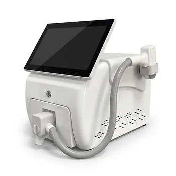
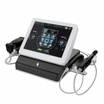
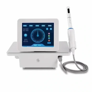
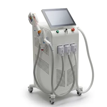
IPL and 1064-nm long-pulsed Nd:YAG systems can help manage persistent vascular redness, telangiectasia, superficial discoloration, and selected vascularized inflammatory lesions after filler injections. IPL uses filtered broadband light to heat superficial hemoglobin and melanin, making it useful for diffuse erythema, fine telangiectasia, and broad areas of uneven pigmentation. The 1064-nm long-pulsed Nd:YAG laser penetrates more deeply and delivers focused thermal energy to larger or deeper vessels and selected inflammatory, granulomatous lesions.
The treatment choice depends on the lesion’s depth, vascular character, and urgency. IPL is generally suited to broad, superficial redness and mixed vascular-pigmentary changes, while long-pulsed 1064-nm Nd:YAG is better suited to deeper or more substantial vascular targets. Neither device should delay urgent evaluation of suspected vascular occlusion, tissue ischemia, infection, or other serious filler complications.
Identifying the Complication Before Treating It
Persistent Changes After the Injection
Swelling and bruising commonly improve within days. A vascular or pigmentary abnormality that persists beyond approximately two weeks may reflect superficial placement, prolonged inflammation, localized vascular congestion, post-inflammatory discoloration, or another filler-related reaction.
The diagnosis should be established before selecting a light-based treatment. The same visible finding can have different causes and therefore require different management.
Telangiectasia and Diffuse Redness
Fine telangiectasia and persistent post-injection erythema are primarily superficial vascular problems. Their principal chromophore is hemoglobin within dilated cutaneous vessels.
IPL can address these changes across relatively broad areas. A vascular laser may be preferable when the vessels are discrete, resistant, or require highly selective treatment.
Blue Discoloration
Blue or blue-gray discoloration after filler may be associated with superficial product placement, visible vascular congestion, or a Tyndall-type optical effect. It should not automatically be treated as a simple pigment lesion.
A careful examination is important because blue discoloration accompanied by pain, blanching, mottling, cool skin, delayed capillary refill, or tissue breakdown may indicate a vascular emergency rather than a cosmetic discoloration.
Vascularized Granulomatous Lesions
Some delayed inflammatory nodules or granulomas may develop associated vascularity and increased local blood supply. Light-based treatment may be considered as part of a broader management plan, particularly when the lesion is superficial or visibly vascular.
The evidence and mechanism are more limited here than for telangiectasia. IPL or Nd:YAG treatment should not be presented as a universal treatment for filler granulomas, which may also require anti-inflammatory, antimicrobial, filler-dissolving, surgical, or other specialist interventions depending on the diagnosis.
How IPL Is Applied
Treating Superficial Vascularity
IPL produces non-coherent broadband light, commonly within a range of approximately 515 to 1200 nm, with cut-off filters selecting the clinically useful spectrum. Filters and treatment parameters are chosen to concentrate energy on superficial hemoglobin while limiting injury to surrounding skin.
Through selective photothermolysis, absorbed light is converted into heat. This can reduce fine telangiectasia, superficial vascular congestion, and diffuse red or pink discoloration.
Managing Mixed Redness and Pigmentation
Post-injection skin changes may combine erythema, superficial vascular dilation, and epidermal pigmentation. IPL can address more than one chromophore because its broadband output can interact with both hemoglobin and melanin.
This makes IPL useful when the treatment area is broad or when the patient has generalized tone irregularity rather than one sharply defined vessel. It is a practical multi-purpose option for clinics that need to treat both vascular and pigmentary findings.
Supporting Inflammatory Lesion Management
By reducing vascularity and local blood supply, IPL may help soften tissue and reduce the apparent volume of selected vascularized inflammatory lesions. This is an adjunctive role rather than a replacement for diagnosing and treating the underlying inflammatory or foreign-body process.
Some patients may also receive intradermal corticosteroid injections as part of the management plan. Combining treatments requires careful clinical judgment because excessive inflammation, infection, and delayed healing must be excluded first.
Where IPL Has the Greatest Practical Value
IPL is most useful for superficial, diffuse, and relatively broad changes, including:
- Fine telangiectasia
- Persistent superficial erythema
- Pink or red discoloration
- Mild superficial vascular congestion
- Mixed vascular and epidermal pigmentation over a broader area
It is less suitable when a lesion is deep, thick, highly localized, or anatomically difficult to access with a contact handpiece.
How 1064-nm Long-Pulsed Nd:YAG Is Applied
Reaching Deeper Vascular Targets
The 1064-nm wavelength penetrates more deeply than the superficial spectra commonly used with IPL. Long pulses deliver thermal energy over a controlled duration, allowing the clinician to target larger or deeper vascular structures.
This makes the system useful for prominent telangiectasia, deeper vascular congestion, and lesions that respond incompletely to superficial treatment.
Treating Larger or Resistant Lesions
Dedicated vascular lasers generally provide greater wavelength specificity and beam control than IPL. A long-pulsed 1064-nm Nd:YAG system may therefore be selected when the vascular target is substantial, resistant, or located deeper in the dermis.
The goal is controlled vascular photothermolysis: energy is absorbed by the target, converted to heat, and used to damage the unwanted vessel while preserving surrounding tissue as much as possible.
Addressing Selected Granulomatous Lesions
The primary reference identifies long-pulsed Nd:YAG as a non-invasive option for larger inflammatory granulomatous lesions. Clinically, this should be understood as treatment of the lesion’s vascular and thermal component, not proof that the laser removes every type of filler granuloma.
A nodule that is firm, painful, enlarging, infected, or diagnostically uncertain requires assessment before laser treatment. Laser therapy may be one component of management when the lesion is appropriately characterized and visibly vascularized.
Anatomical Precision
Unlike IPL handpieces, dedicated lasers deliver a focused, collimated beam. This allows more precise application to discrete vessels and can be advantageous on facial areas where treatment must be carefully confined.
The clinician must still account for the 1064-nm wavelength’s deeper tissue penetration and the risk of unwanted thermal injury, especially in thin skin or near sensitive structures.
Choosing Between IPL and Nd:YAG
Choose IPL for Broad Superficial Change
IPL is generally favored when the dominant problem is diffuse superficial redness, fine telangiectasia, or combined vascular and pigmentary discoloration across a relatively large area.
Its interchangeable filters also make it a versatile platform for clinics treating multiple superficial aesthetic indications.
Choose Nd:YAG for Depth and Selectivity
Long-pulsed 1064-nm Nd:YAG is generally more appropriate for deeper, larger, or discrete vascular targets. It offers greater wavelength specificity and may provide stronger vascular targeting than broadband IPL for resistant lesions.
Its deeper action is an advantage when the clinical target is deep, but it also increases the importance of conservative parameter selection and anatomical expertise.
Consider a Staged or Combined Strategy
Some patients have both focal vascular lesions and diffuse background redness or pigmentation. In such cases, a staged approach may use a precise vascular laser for the main lesion and IPL for broader residual erythema or tone irregularity.
Treatment should be individualized rather than automatically combined. Each additional thermal treatment increases the need to control cumulative inflammation and protect the skin barrier.
Understanding the Trade-offs
IPL Offers Versatility but Less Precision
IPL can treat vascular and pigmentary chromophores with one platform, but its broadband output is less selective than a dedicated single-wavelength laser. It is therefore a flexible workhorse rather than the most precise option for every vascular lesion.
The IPL handpiece must contact the skin, and that contact can make immediate visualization of the tissue response more difficult. Precise treatment in concave facial areas may also be challenging.
Nd:YAG Offers Depth but Requires Greater Control
The 1064-nm Nd:YAG laser can reach deeper vessels and larger targets, but deeper thermal delivery carries a greater risk of pain, swelling, blistering, pigmentary change, or unintended tissue injury if parameters are poorly matched.
Long-pulsed Nd:YAG is also not a universal solution for superficial discoloration. A superficial pink lesion may be better addressed with IPL or another wavelength selected specifically for its vascular depth and caliber.
Light Treatment Does Not Correct Every Filler Reaction
Vascular lasers and IPL can manage visible vascular sequelae, but they do not independently resolve every cause of post-filler discoloration or nodularity. Persistent filler material, infection, delayed hypersensitivity, foreign-body inflammation, and true granuloma formation may need other treatments.
In particular, suspected vascular occlusion is an urgent clinical problem. Light-based treatment should not substitute for immediate vascular assessment and established emergency management.
Adverse Effects Need Anticipation
Possible treatment effects include temporary redness, swelling, crusting, discomfort, pigmentary alteration, and thermal injury. Risk depends on skin type, treatment settings, lesion depth, recent inflammation, tanning, and the proximity of sensitive facial structures.
A conservative test area, appropriate cooling, eye protection, and careful follow-up are important components of responsible treatment. The exact settings should be determined by a qualified clinician rather than copied across devices or patients.
Making the Right Choice for Your Goal
The correct device is selected after confirming that the problem is vascular and that urgent complications have been excluded.
- If your primary focus is diffuse superficial redness or fine telangiectasia: Consider filtered IPL when a broad treatment area or combined vascular-pigmentary correction is needed.
- If your primary focus is a discrete, deeper, larger, or resistant vascular lesion: Consider a long-pulsed 1064-nm Nd:YAG system for more selective and deeper thermal targeting.
- If your primary focus is a vascularized inflammatory nodule or granuloma: Use IPL or Nd:YAG only as an appropriately selected adjunct after diagnostic assessment and consideration of anti-inflammatory or other treatment.
- If your primary focus is blue discoloration with pain, mottling, blanching, or poor perfusion: Seek urgent vascular evaluation rather than treating the appearance with IPL or laser.
- If your primary focus is broad skin-tone recovery after uncomplicated inflammation: IPL may help residual superficial vascularity and pigmentation, provided active infection and significant ongoing inflammation have been excluded.
The safest clinical strategy is to match the device to the depth and cause of the lesion, while treating urgent vascular complications as emergencies rather than cosmetic discolorations.
Summary Table:
| Feature | IPL | 1064-nm Long-Pulsed Nd:YAG |
|---|---|---|
| Wavelength | Broadband (515-1200 nm) | 1064 nm (single wavelength) |
| Depth | Superficial | Deep |
| Optimal Targets | Fine telangiectasia, diffuse erythema, mixed pigment | Larger/deeper vessels, resistant lesions |
| Selectivity | Lower | Higher |
| Common Uses | Broad superficial redness, mixed vascular-pigmentary changes | Deep vascular lesions, vascularized granulomas |
Elevate your clinic's filler complication management with BELIS professional-grade IPL and Nd:YAG systems. Our advanced technology offers precise, effective solutions for vascular and pigmentary issues. Partner with us to enhance your aesthetic practice. Contact us today to learn more!
Related Products
- Clinic Use IPL SHR ND YAG Laser Hair Removal RF Skin Tightening Machine
- Clinic Use IPL and SHR Hair Removal Machine with Nd Yag Laser Tattoo Removal
- Q Switch Nd Yag Laser Machine Tattoo Removal Nd Yag Machine
- Tri Laser Diode Hair Removal Machine Professional Beauty Equipment
- 9D 7D HIFU Vaginal RF Lifting Treatment
People Also Ask
- Why is the millisecond pulse width critical for laser hair removal on dark skin? Safety Guide for Dark Skin Treatments
- How does the etiology of Pseudofolliculitis Barbae (PFB) explain why traditional shaving fails, and how can professional hair removal lasers provide a superior clinical solution? Discover the Ultimate Treatment
- How does the combined application of laser devices and Eflornithine cream create a synergistic effect? Maximize Results
- What are the core hardware limitations of home hair removal devices? Why Professional Systems Deliver Superior Results
- What research methods and biological markers are used to evaluate the efficacy and penetration depth of hair removal lasers? Explore key techniques and assays.