Pre-treatment phototesting and skin phototype evaluation are essential because they reveal how an individual’s skin is likely to react before high-energy light is applied. This information helps clinicians select appropriate fluence, pulse duration, wavelength, and optical filters for lasers or phototherapy. It reduces the risk of excessive heating, phototoxic erythema, burns, delayed healing, and post-inflammatory hyperpigmentation while improving the likelihood of an effective result.
Skin type and photoreactivity determine how safely skin can absorb light energy. Combining phototype classification with objective skin assessment and, when appropriate, a test spot allows treatment parameters to be individualized rather than applied according to a generalized protocol.
Why Skin Reactivity Must Be Assessed First
Skin Does Not Absorb Light Uniformly
Epidermal melanin competes with the intended treatment target for absorbed light energy. Patients with higher melanin levels, particularly Fitzpatrick Types IV-VI, may absorb more energy near the skin surface and experience greater thermal injury risk.
A phototype evaluation estimates constitutional pigmentation and the person’s typical tendency to burn or tan. Objective diagnostic devices can add information about melanin density, erythema response, hydration, barrier condition, and sensitivity.
A Test Spot Shows Individual Response
A pre-treatment test spot provides practical evidence of how the patient responds to a selected wavelength and energy level. It can reveal excessive erythema, blistering, crusting, or abnormal pigmentation before the full treatment area is exposed.
This is especially important when treating new patients, darker phototypes, recently tanned skin, or areas with uncertain sensitivity.
Treatment Parameters Must Be Customized
Phototype and phototesting findings guide decisions about energy fluence, pulse duration, wavelength, spot size, and cutoff filters. Longer wavelengths or appropriate filters may help reduce superficial melanin absorption while allowing energy to reach the intended target.
The correct setting is not simply the highest setting a device can deliver. It is the lowest effective level that achieves the clinical objective without causing unacceptable tissue injury.
What the Evaluation Can Identify
Baseline Pigment and Erythema
Measuring baseline pigmentation and erythema helps distinguish a normal treatment response from a pre-existing condition. It also provides a reference for monitoring changes after treatment.
Without a baseline, persistent redness or darkening may be difficult to interpret accurately.
Barrier Damage and Inflammation
Skin analysis can identify compromised barrier function, dehydration, active inflammation, or increased sensitivity. Applying intense light to already-irritated skin can increase the risk of severe erythema, dermatitis, delayed healing, and pigmentary complications.
The procedure may need to be postponed until the barrier or inflammatory condition has been addressed.
Photosensitizing Conditions and Medications
Some patients have photodermatoses, active infections, or medical conditions that make light exposure unsafe. Photosensitizing medications can also increase the likelihood of an exaggerated reaction.
A proper consultation should therefore include relevant medical history, current medications, recent sun exposure, tanning, and previous reactions to light-based treatments.
Suspicious Lesions and High-Pigment Areas
Visual examination may identify suspicious pigmented lesions or areas where unusually high melanin content could absorb excessive energy. Uncertain lesions should be evaluated appropriately before treatment rather than treated cosmetically without clarification.
This assessment protects the patient and prevents light from being applied to an area requiring medical diagnosis.
How Phototype Affects Laser and Phototherapy Decisions
Fitzpatrick Types I-III
Lighter phototypes generally contain less epidermal melanin and may have a lower risk of superficial melanin-related heat absorption. They can still develop burns, erythema, scarring, or pigmentary changes if energy is excessive or the skin is inflamed.
Phototype classification reduces risk, but it does not replace test spots, clinical judgment, or conservative parameter selection.
Fitzpatrick Types IV-VI
Darker phototypes require particular attention because epidermal melanin can absorb more of the delivered energy. This increases the risk of overheating and post-inflammatory hyperpigmentation.
Appropriate wavelength selection, longer pulse strategies where indicated, suitable filters, cooling, conservative starting parameters, and test spots can improve the safety margin.
Phototype Is Only One Variable
The Fitzpatrick scale is based partly on reported sun response and is not a complete measurement of treatment risk. Two patients with the same classification can differ in melanin distribution, tanning status, barrier health, medication exposure, and history of hyperpigmentation.
For that reason, phototype assessment should be combined with direct skin examination and objective diagnostic information where available.
Understanding the Trade-offs
Conservative Settings May Require More Sessions
Lower initial energy or more cautious treatment parameters may reduce complications but may also produce slower or less dramatic results. The clinician may need to increase treatment gradually or schedule additional sessions.
This is an acceptable trade-off when the alternative is causing an injury that can be difficult to reverse.
A Test Spot Cannot Eliminate All Risk
A test spot samples only a small area and may not predict every delayed response. Hyperpigmentation, prolonged erythema, or other complications can appear after the test period or behave differently across anatomical sites.
The test spot is a risk-reduction tool, not a guarantee of safety.
Device Settings Cannot Be Transferred Uncritically
A setting that worked for one patient, device, body area, or indication may be inappropriate for another. Device architecture, wavelength, pulse profile, cooling system, treatment target, and operator technique all affect tissue response.
Clinical protocols must therefore be individualized rather than copied from a generic chart.
Delaying Treatment May Be the Correct Decision
Recent tanning, active infection, uncontrolled inflammation, photosensitizing medication, or suspicious lesions may make immediate treatment inappropriate. Postponement or referral is sometimes the safest and most clinically responsible outcome.
Making the Right Choice for Your Goal
A structured evaluation should connect the patient’s goal with their actual risk profile before energy is delivered.
- If your primary focus is patient safety: Complete a medical and skin-history review, assess barrier and inflammatory status, classify phototype, and use a test spot when clinically appropriate.
- If your primary focus is preventing hyperpigmentation: Pay particular attention to darker phototypes, recent tanning, baseline pigment, previous pigmentary reactions, conservative fluence, suitable wavelengths, and post-treatment monitoring.
- If your primary focus is treatment efficacy: Match wavelength, pulse duration, fluence, filters, and cooling to the intended chromophore and the patient’s measured skin characteristics.
- If your primary focus is identifying contraindications: Screen for photosensitizing medications, photodermatoses, active infections, suspicious pigmented lesions, and other conditions that require postponement or medical evaluation.
The safest high-energy light treatment begins with measuring the skin’s capacity to tolerate it, not with selecting a device setting.
Summary Table:
| Why Phototesting & Phototype Evaluation? | How It Helps |
|---|---|
| Skin does not absorb light uniformly | Identifies melanin-related heat risk, guides energy selection |
| A test spot shows individual response | Reveals adverse reactions before full-area treatment |
| Treatment parameters must be customized | Allows tailoring fluence, pulse duration, and wavelength |
| Detects baseline pigment and erythema | Provides a reference for monitoring post-treatment changes |
| Identifies barrier damage or inflammation | Prevents worsening of sensitive or compromised skin |
| Screens photosensitizing conditions | Avoids unsafe light exposure in at-risk patients |
| Discovers suspicious lesions | Prevents inadvertent treatment of undiagnosed conditions |
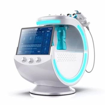
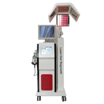
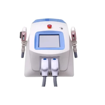
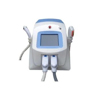
Ensure safe, effective high-energy light treatments with BELIS's advanced diagnostic and aesthetic devices. Our professional-grade lasers (Diode, Alexandrite, CO2, Erbium, Nd:YAG, Pico), IPL, and PDT systems are designed for clinics and premium salons. Partner with us to enhance patient safety and treatment outcomes. Contact us today to discuss how our technology can elevate your practice.
Related Products
- Hydrafacial Machine with Facial Skin Analyzer Skin Tester
- Skin Tester Analysis Machine Analyser for Skin Testing
- Hydrofacial Machine with Facial Skin Analyzer and Skin Tester
- Skin Tester Analysis Machine Analyser for Skin Testing
- 22D HIFU Machine Device Facial Machine
People Also Ask
- What is the difference between HydraFacial and hydrodermabrasion? Choose the Right Facial for Your Skin
- How does the HydraFacial machine work? Master the 4-in-1 Vortex Technology for Flawless Skin
- What are the general outcomes and benefits of a HydraFacial treatment? Achieve Instant Glow and Skin Health
- Who can use a HydraFacial machine? A Guide to Legal and Safe Operation
- What does a HydraFacial machine do? Achieve Radiant Skin with No Downtime