The specific window of 2 to 6 months post-procedure is critical because it represents the "pathological evolution phase" of delayed adverse reactions, specifically Fox-Fordyce disease. Unlike immediate side effects like burns or erythema, this condition requires time for damaged follicular structures to undergo physiological changes. Monitoring during this timeframe is the only way to detect the gradual formation of keratotic plugs and the resulting chronic inflammation.
The 2 to 6-month period is when microscopic damage to the follicular infundibulum matures into visible clinical lesions. Clinical observation is vital during this phase to identify apocrine secretion obstruction that was triggered by the initial laser treatment but remained asymptomatic during the early healing stages.
The Mechanism of Delayed Onset
The Pathological Evolution Phase
Laser hair removal works by thermally damaging the hair follicle. However, in specific cases leading to Fox-Fordyce disease, this damage sets off a slow-moving chain reaction.
The skin often appears to heal normally immediately after the procedure. The critical changes are happening beneath the surface, taking months to manifest visually.
Structural Damage to the Infundibulum
The primary reference indicates that the laser causes specific damage to the follicular infundibulum. This is the funnel-shaped upper portion of the hair follicle.
Over the course of several months, this damage alters how the follicle sheds skin cells.
Formation of Keratotic Plugs
As the infundibulum attempts to heal or react to the damage, it begins to produce excess keratin.
This leads to the gradual formation of "keratotic plugs." These are physical blockages that seal off the opening of the follicle.
Consequences of Follicular Obstruction
Blocking Apocrine Secretion
Once the keratotic plugs have fully formed, they obstruct the exit path for apocrine sweat glands.
This entrapment of secretions is the direct cause of the lesions associated with Fox-Fordyce disease. It does not happen immediately; it requires the plug to fully mature first.
The Onset of Chronic Inflammation
The trapped secretions eventually rupture the follicular wall or cause significant pressure.
This triggers a localized immune response, resulting in chronic inflammation. This inflammation is what clinicians finally observe as lesions between the 2 and 6-month marks.
Common Pitfalls in Follow-Up Protocols
The Risk of Premature Discharge
A significant clinical error is terminating patient follow-up after visible surface healing (typically 2-4 weeks).
Ending care this early guarantees that conditions with delayed onsets, such as Fox-Fordyce disease, will be missed entirely or misdiagnosed by other providers later.
Misinterpreting the Timeline
Because the reaction occurs months after the "injury," patients and clinicians may fail to link the symptoms to the laser procedure.
Without knowledge of this 2-6 month evolution window, the inflammation may be mistreated as a new, unrelated dermatological issue rather than a sequela of the laser treatment.
Integrating This into Clinical Practice
To ensure comprehensive patient care, follow-up protocols must account for the biological timeline of follicular pathology.
- If your primary focus is Early Detection: Schedule a specific "delayed onset check-up" at the 3 or 4-month mark to inspect for signs of follicular plugging.
- If your primary focus is Differential Diagnosis: Evaluate any new inflammatory lesions appearing in this window specifically for Fox-Fordyce characteristics rather than common folliculitis.
True clinical vigilance requires monitoring not just for immediate injury, but for the long-term physiological evolution of the treated tissue.
Summary Table:
| Phase | Timeframe | Pathological Evolution | Clinical Observation Focus |
|---|---|---|---|
| Immediate Healing | 0 - 4 Weeks | Surface repair, erythema reduction | Acute side effects (burns, redness) |
| Pathological Evolution | 2 - 6 Months | Keratotic plug formation & apocrine blockage | Delayed reactions (Fox-Fordyce disease) |
| Chronic Response | 6+ Months | Persistent inflammation & lesion maturity | Long-term sequela management |
Elevate Your Clinic’s Safety with BELIS Technology
Ensure the highest standards of patient care and long-term safety with BELIS. As specialists in professional-grade medical aesthetic equipment for clinics and premium salons, we provide the precision tools you need to minimize risks and maximize results. Our advanced laser systems (Diode Hair Removal, CO2 Fractional, Nd:YAG, Pico), alongside HIFU, Microneedle RF, and body sculpting solutions like EMSlim and Cryolipolysis, are engineered for clinical excellence.
Partner with BELIS to access cutting-edge technology and expert support that helps you navigate complex clinical timelines with confidence. Contact us today to discover how our equipment can enhance your practice’s reputation and patient outcomes.
References
- Rita Sammour, Constantin El Habr. Fox–Fordyce Disease: An under‐diagnosed adverse event of laser hair removal?. DOI: 10.1111/jdv.13680
This article is also based on technical information from Belislaser Knowledge Base .
Related Products
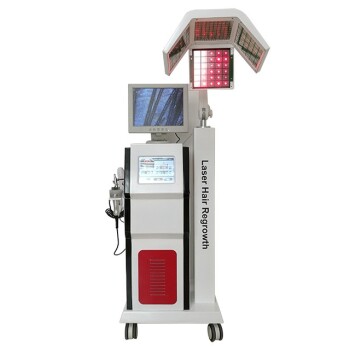
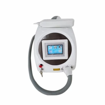
- Diode Tri Laser Hair Removal Machine for Clinic Use
- Diode Laser SHR Trilaser Hair Removal Machine for Clinic Use
- Clinic Diode Laser Hair Removal Machine with SHR and Trilaser Technology
- Clinic Use IPL SHR ND YAG Laser Hair Removal RF Skin Tightening Machine
- Trilaser Diode Hair Removal Machine for Beauty Clinic Use
People Also Ask
- Why can traditional high-energy pulse (HR) modes lead to adverse skin reactions? Understanding Thermal Injury Risks
- What is the technical significance of adjusting the pulse duration in the Diode Laser hair removal process?
- What are the advantages of the Constant Motion Technique? Elevate Your Clinic's Laser Hair Removal Results
- What is the working mechanism of professional-grade Diode laser equipment? Master the Physics of Selective Photothermolysis
- What is the primary mechanism of action for Diode Laser hair removal? Mastering Selective Photothermolysis