The key predictors are location, size, depth, vessel characteristics, color, and patient age. Lesions on the face, neck, trunk, chest, and upper arms generally respond better than lesions on the hands or legs; lateral facial and forehead regions often outperform central mid-face areas. Smaller, flat, pink lesions are usually more responsive than large, thickened, dark-purple, nodular, or high-flow lesions, which may require deeper-penetrating wavelengths and more cautious parameter selection.
Port Wine Stain outcomes depend on matching energy delivery to vessel anatomy. Practitioners should assess where the lesion is, how extensive and thick it is, how deep and ectatic its vessels appear, and whether age-related hypertrophy or high-flow features are present before selecting wavelength, pulse duration, fluence, spot size, and cooling.
Start With the Lesion’s Anatomy
Anatomical location predicts clearance
Port Wine Stains on the face, neck, trunk, chest, and upper arms generally demonstrate better clearance than lesions on distal extremities such as the hands and legs.
Within the face, lateral facial regions and the forehead tend to respond better than central mid-facial areas. The V3 lateral facial region is generally more favorable than the central V2 distribution.
These differences should influence counseling. The same laser settings should not be expected to produce equivalent results across every body site.
Lesion size affects treatment efficiency
Lesions smaller than approximately 20 cm² generally clear more rapidly and completely than extensive lesions.
Large lesions may require more treatment sessions and create greater difficulty in maintaining consistent spot placement, cooling, and energy delivery across the entire area.
Size is therefore both a biological and procedural consideration: extensive disease may be less responsive and more challenging to treat uniformly.
Vessel depth determines the required penetration
Histologically, PWS vessels are concentrated predominantly within the upper 0.6 mm of the dermis, but their depth, diameter, and degree of ectasia vary with lesion maturity.
Flat, superficial lesions with small vessels may respond well to dedicated vascular lasers or other light-based systems. Thicker lesions with deeper or more ectatic vessels may require a wavelength and pulse strategy capable of delivering energy farther into the dermis, such as a long-pulsed Nd:YAG or Alexandrite approach when clinically appropriate.
The objective is not simply to use more energy. It is to deliver sufficient thermal energy to intravascular hemoglobin while preserving the epidermis and surrounding dermal structures.
Evaluate Color, Thickness, and Vascular Behavior
Color reflects lesion maturity
Early or superficial PWS lesions are often pale pink and relatively flat. These lesions commonly provide more favorable treatment responses.
With time, vessels may progressively dilate, increasing the concentration of erythrocytes and shifting the lesion toward dark purple. Darker lesions are often more resistant and require particular caution because excessive energy density can increase epidermal heating and complications.
Color should therefore guide, but not independently determine, fluence selection. It must be interpreted alongside vessel depth, thickness, patient age, and treatment response.
Thickening and nodularity predict poorer outcomes
A lesion that is hypertrophic, thickened, or nodular at presentation is less likely to clear as readily as a flat lesion.
These clinical features suggest more advanced vascular remodeling and potentially deeper or larger-caliber vessels. They may require a different treatment strategy, more sessions, or realistic counseling that complete clearance may be difficult.
Dermoscopic features can refine the assessment
Dermoscopic evidence of deep, thick, high-flow blood vessels in the subpapillary plexus is associated with poorer outcomes.
This information can help explain why two lesions with similar surface color may respond differently. It also supports a more cautious approach to wavelength, pulse duration, fluence, and the expected number of sessions.
Account for Patient Age and Lesion Evolution
Earlier treatment may improve the prognosis
Treatment during early childhood, ideally beginning around one to two years of age when clinically appropriate, is associated with higher clearance rates.
The rationale is that vessels may undergo progressive ectasia and hypertrophy with age. Treating before these changes become pronounced may improve responsiveness.
Age also affects practical treatment decisions. Pediatric patients may require lower starting fluences, topical anesthesia, sedation, or general anesthesia, together with complete ocular protection when treating near the eyes.
Age changes both biology and tolerability
A younger patient may have a more favorable lesion biology but may be less able to tolerate repeated procedures while awake.
Conversely, an older patient may tolerate treatment more readily but present with thicker, darker, and more resistant vessels. The treatment plan must therefore balance expected biological response with anesthesia, safety, and procedural feasibility.
Translate the Assessment Into Laser Settings
Choose wavelength according to vessel depth
For superficial, flat lesions, dedicated vascular systems targeting hemoglobin may be appropriate.
For thicker or deeper lesions, deeper-penetrating modalities such as long-pulsed Nd:YAG or Alexandrite lasers may be considered. The choice should follow the suspected vessel depth and clinical morphology rather than relying on a single standard protocol.
Adjust pulse duration to vessel size
Pulse duration should be selected in relation to the thermal behavior and size of the target vessels.
Superficial lesions with smaller vessels may be treated using shorter vascular-laser pulses, while thicker or deeper vessels may require a pulse strategy that allows adequate thermal delivery without excessive injury to surrounding tissue.
The correct pulse duration is therefore part of a coordinated decision with wavelength, spot size, fluence, and cooling.
Use fluence cautiously and progressively
Fluence determines the thermal energy delivered per unit area. Initial settings should be individualized using test patches, particularly when lesion color, thickness, anatomical sensitivity, or patient age increases risk.
Reference protocols for vascular laser systems may use fluences in ranges such as 4.5–8 J/cm² with smaller spots, while other dedicated 595 nm protocols use approximately 6.5–7.0 J/cm² with a larger spot. These figures are equipment- and patient-dependent starting frameworks, not universal prescriptions.
Fluence may be increased gradually as the lesion lightens, commonly in increments of approximately 0.25–0.5 J/cm² when clinically justified. Non-responding areas should not simply be repeatedly overtreated.
Use spot size and cooling to control heat distribution
Larger spot sizes can support more uniform and deeper heat distribution, while smaller spots may be useful for localized work or particular device configurations.
Continuous external or integrated cooling helps protect the epidermis and permits safer energy delivery. Cooling is especially important when treating darker lesions or anatomically sensitive areas.
Interpret the immediate endpoint correctly
With pulsed dye laser treatment, the intended endpoint is typically purpura, reflecting vascular injury and photothermolysis.
A purplish-gray or dark violet response can indicate effective treatment, but the endpoint must be interpreted in the context of the device and protocol. Failure to see an immediate response does not justify repeated pulses to the same site during that session.
Use a Structured Treatment-Response Process
Test before committing to broad treatment
Fluence test patches allow the practitioner to evaluate both efficacy and adverse effects before treating the entire lesion.
The response should be assessed after adequate healing and, commonly, at approximately six to eight weeks. This delayed evaluation is more informative than relying only on the immediate appearance.
Space treatments to allow biological response
Typical vascular laser protocols may involve three to four sessions spaced approximately three months apart, although the actual schedule depends on healing, response, device, and lesion characteristics.
Pulsed-light approaches for milder or diffuse lesions may use different intervals, such as approximately 40 days. Treatment timing should not be standardized without considering the modality and tissue response.
Avoid cumulative thermal injury
Spot placement should be contiguous, with only limited overlap. The references describe slight overlap—approximately 5–10% in one delivery approach—and approximately 25% in a specific focusing-handpiece protocol.
Because overlap recommendations vary by device and handpiece, practitioners should follow the validated manufacturer protocol for the equipment being used. Large overlaps and repeated pulses over the same site increase localized heat accumulation and the risk of epidermal damage, hyperpigmentation, and scarring.
Understanding the Trade-offs
More energy is not automatically better
Increasing fluence may improve vessel destruction, but it also increases the risk of epidermal injury, post-inflammatory hyperpigmentation, and scarring.
The appropriate goal is the lowest effective energy that achieves the intended vascular endpoint, followed by reassessment rather than immediate escalation.
A darker lesion may need caution, not aggressive treatment
Dark-purple lesions can contain more blood and may appear to be obvious targets, but they are also frequently more resistant and may absorb substantial energy.
Using excessive power density can overheat the epidermis. Darker lesions therefore require careful test spots, cooling, conservative escalation, and realistic expectations.
Deeper treatment can increase collateral risk
A deeper-penetrating wavelength may be useful for thick or nodular lesions, but deeper energy delivery can also increase the risk of damaging non-target structures.
The practitioner must balance penetration against tissue safety and avoid assuming that a deeper modality will guarantee complete clearance.
Immediate appearance does not determine final clearance
Purpura is a useful immediate endpoint for some vascular laser systems, but it is not equivalent to complete long-term clearance.
Final assessment requires healing and follow-up. Multiple sessions are often necessary, particularly for large, mature, thick, or anatomically unfavorable lesions.
How to Apply This to Your Project
A defensible treatment plan should document the lesion’s anatomy, morphology, expected response, and safety limits before settings are chosen.
- If your primary focus is maximizing clearance: Prioritize early, flat, superficial lesions in favorable locations and match wavelength and pulse duration to the observed vessel depth.
- If your primary focus is treating thick or dark-purple lesions: Assess for deeper, ectatic, nodular, or high-flow vessels and consider a deeper-penetrating strategy with conservative test patches.
- If your primary focus is minimizing complications: Use individualized starting fluence, effective cooling, careful spot overlap, and avoid repeated pulses or aggressive escalation in non-responding areas.
- If your primary focus is counseling patients: Explain that location, lesion size, age, color, thickness, and vascular architecture affect the number of sessions and the likelihood of complete clearance.
The safest and most predictive approach is to treat the lesion’s vascular anatomy—not merely its visible color—with carefully tested, progressively optimized settings.
Summary Table:
| Factor | Impact on Treatment |
|---|---|
| Location | Face, neck, trunk, upper arms respond better than extremities; lateral face better than central. |
| Size | Smaller (<20 cm²) clear faster; larger lesions need more sessions and careful technique. |
| Vessel Depth | Superficial vessels respond to vascular lasers; deeper vessels may need Nd:YAG or Alexandrite. |
| Color | Pale pink responds well; dark purple is more resistant and requires caution. |
| Thickness | Flat lesions clear better; thickened or nodular lesions have poorer outcomes. |
| Age | Early treatment (1-2 years) improves prognosis; age affects tolerability and vessel maturity. |
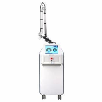
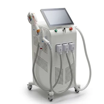
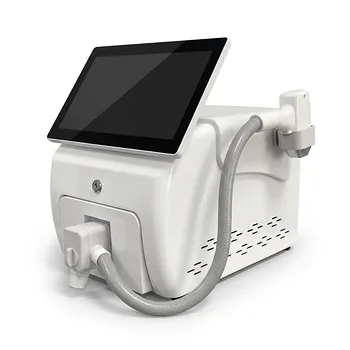
Optimize your PWS laser treatments with BELIS's advanced aesthetic devices. Our portfolio includes precise vascular lasers and IPL systems designed for superior outcomes. Contact our experts today to enhance your practice and patient satisfaction!
Related Products
- Hydrofacial Machine with Facial Skin Analyzer and Skin Tester
- Pico Laser Tattoo Removal Machine Picosure Picosecond Laser Machine
- Clinic Use IPL and SHR Hair Removal Machine with Nd Yag Laser Tattoo Removal
- 808nm Diode Laser Hair Removal Machine 755+808+1064nm Mixed Wavelength Professional Equipment
- Clinic Use IPL SHR ND YAG Laser Hair Removal RF Skin Tightening Machine
People Also Ask
- What is a HydroFacial treatment? Discover the Secret to Instant, Radiant Skin with Vortex-Fusion Technology
- What is the first step of a hydro-facial treatment? Master the Art of Professional Cleansing & Exfoliation
- What is a Hydrafacial machine and what are its unique features? Elevate Your Clinic with Vortex-Fusion Tech
- What are the business benefits of a spa or salon owning a Hydrafacial machine? Drive Revenue and Loyalty
- Is a HydraFacial machine worth it? Achieve Instant Glow with No Downtime