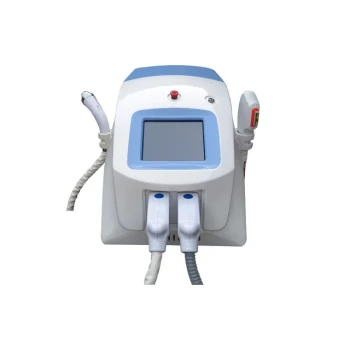
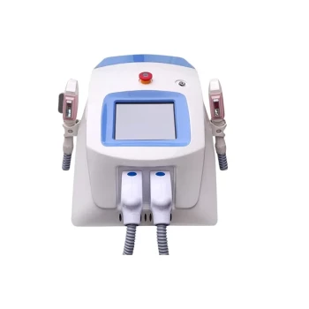
Unprotected exposure to high-intensity light radiation can injure both the ocular surface and deeper eye structures. Acute complications include phototoxic keratitis, conjunctivitis, corneal irritation, and thermal injury. Depending on the wavelength, intensity, exposure time, and treatment location, radiation can also cause retinal photochemical damage, macular injury, and damage to the iris, uvea, or choroid. Clinics should therefore treat wavelength-specific eye protection as a mandatory control for every laser, IPL, phototherapy, LED, and other high-intensity light procedure.
The primary safety principle is simple: control the beam, protect every exposed eye, and use protection rated for the device’s exact wavelength and operating conditions. Standard protective eyewear may be insufficient during periorbital treatment, where dedicated opaque or intraocular shields may be required.
How High-Intensity Light Injures the Eye
Ocular Surface Injury
UV and intense visible or infrared radiation can irritate or damage the cornea and conjunctiva. Possible acute effects include phototoxic keratitis, conjunctivitis, pain, redness, tearing, light sensitivity, and corneal epithelial injury.
These injuries may occur even when exposure is brief, particularly when the source is close to the face or the radiation exceeds applicable exposure limits.
Retinal and Macular Injury
Visible light and near-infrared radiation can be focused by the eye onto the retina. High-intensity exposure may produce retinal burns or photochemical injury, with the macula at particular risk because of its role in central vision.
Blue light in approximately the 400–450 nm range presents a significant photochemical hazard. Because near-infrared radiation is invisible, it may be especially dangerous: it does not reliably trigger the normal blink or aversion response.
Deeper Ocular Damage
Depending on wavelength and exposure conditions, excessive radiation can injure the iris, uvea, choroid, or other intraocular tissues. Thermal injury is a particular concern with high-energy IPL and laser systems, especially when treatment is performed near the orbit.
The severity of injury depends on wavelength, fluence, pulse duration, beam geometry, distance, exposure time, and whether the beam is direct, reflected, or diffuse.
Which Procedures Require Particular Caution?
IPL Treatments
IPL emits intense, polychromatic light rather than a single laser wavelength. Its broad spectrum and deep penetration create risks from both ocular absorption and thermal effects.
Risk increases during facial or periorbital treatment, when light can enter through the eyelids or reach the eye through gaps in ordinary protective equipment.
Laser Procedures
Diode, Alexandrite, Nd:YAG, and other medical lasers can cause serious ocular injury through direct beams, specular reflections, or diffuse scatter. The appropriate eyewear must match the active wavelength and provide sufficient optical density for the device’s output.
The applicator should not emit until it is correctly positioned. Reflective instruments, jewelry, and other shiny objects should be removed or controlled around the beam path.
Phototherapy and LED Systems
LED systems are generally less hazardous than surgical lasers because they emit non-coherent light. They are not automatically risk-free, however: visible and near-infrared output can still exceed ocular exposure limits and focus on the retina.
UV phototherapy also requires dedicated eye protection because UV exposure can damage the ocular surface and other eye tissues. NIR exposure requires additional caution because it is invisible.
How Clinics Should Implement Safety Protocols
Use Wavelength-Specific Protective Eyewear
Both patients and all personnel present should wear certified protective eyewear appropriate to the emitted wavelength or wavelength range. Protection should be selected using the device manufacturer’s requirements, applicable exposure limits, and the required optical density.
Generic goggles are not an adequate substitute when their wavelength range, optical density, or damage threshold has not been verified.
Apply Stronger Protection Near the Orbit
For procedures close to the eye, external goggles may not provide sufficient protection. Clinics should use opaque eye cups or dedicated patient shields for facial treatments and appropriately designed metal intraocular shields when treatment is performed inside the bony orbit.
Intraocular shields must be placed only by appropriately trained clinicians. Patients should remove soft or hard contact lenses before shield placement to reduce the risk of corneal trauma or abrasion.
Protect Non-Target Tissue
Non-target tissue should be shaded or covered whenever practical. During periocular irradiation, patients should keep their eyes closed as instructed, but closed eyelids should not be treated as the primary protective barrier.
For UV and other light therapies, use the dedicated protective glasses or shields specified by the treatment protocol throughout exposure.
Control the Treatment Environment
The treatment room should be arranged to minimize direct and reflected exposure. Clinics should:
- Restrict access during emission.
- Prevent reflective objects from entering the beam path.
- Keep doors closed or use appropriate warning signs and indicators.
- Confirm that the applicator is secure before activating emission.
- Control stray radiation within the operator’s workspace.
- Ensure that protective eyewear is available before treatment begins.
Contact applicators can help reduce hazardous stray reflections in some procedures, but they do not replace appropriate eye protection or room controls.
Verify Device and Eyewear Compatibility
Before each treatment, staff should confirm the device type, active wavelength, treatment parameters, and required protective equipment. Eyewear should be inspected for cracks, scratches, contamination, poor fit, and expired or damaged components.
For LED and NIR systems, protection must be rated for the exact operating spectrum. A visible-light filter may not adequately protect against invisible NIR radiation.
Standardize Staff Training and Documentation
Eye protection should be a mandatory checklist item, not an optional judgment call. Staff training should cover:
- Direct, reflected, and diffuse exposure hazards.
- The limits of ordinary glasses and closed eyelids.
- Correct selection and use of wavelength-specific eyewear.
- Intraocular shield placement and removal, where applicable.
- Emergency shutdown procedures.
- Reporting and assessment of suspected ocular exposure.
The clinic should document device parameters, protective equipment used, staff compliance, and any adverse symptoms or incidents.
Understanding the Trade-offs
Protection Must Match the Procedure
Overly dark or poorly selected eyewear can interfere with communication, positioning, or visualization of the treatment area. The solution is not to reduce protection, but to use certified equipment that provides the required protection while allowing safe clinical workflow.
Cooling Does Not Protect the Eye
Integrated epidermal cooling, cold air, and cryogen systems can reduce skin heating, but they do not prevent retinal or corneal exposure. Cooling devices must also be used carefully near the eye because excessive cooling, dryness, or topical agents can irritate or damage the cornea.
Skin Safety and Eye Safety Are Separate Controls
Darker or tanned skin can absorb more energy during some light-based procedures, increasing the risk of nonspecific thermal injury. Adjusting fluence and pulse duration may protect the skin, but it does not replace ocular controls.
Screening Does Not Replace Engineering Controls
A baseline ophthalmic examination may be appropriate before an extended phototherapy course, with periodic follow-up for long-term treatment. Screening helps identify pre-existing risk and monitor changes, but it cannot make an unsafe exposure acceptable.
Recognizing a Possible Exposure
Symptoms Requiring Prompt Assessment
After a suspected exposure, symptoms such as eye pain, persistent redness, tearing, photophobia, blurred vision, a central blind spot, or new visual distortion require prompt clinical assessment.
Symptoms may not always appear immediately, particularly after retinal photochemical exposure. The exposure should be documented, the device removed from service if a malfunction is suspected, and the patient referred for appropriate ophthalmic evaluation.
Do Not Rely on Symptoms Alone
The absence of immediate pain does not establish safety. Invisible infrared radiation and some retinal injuries may bypass the normal protective blink response and may not produce an obvious early warning.
Making the Right Choice for Your Goal
Use the same controls consistently across every light-based energy procedure, then increase protection when treatment approaches the orbit.
- If your primary focus is patient safety: Use certified wavelength-specific eyewear for every procedure and dedicated opaque or intraocular shields for periorbital treatment when indicated.
- If your primary focus is staff safety: Control direct, reflected, diffuse, and invisible NIR exposure through room access controls, beam-path management, compatible eyewear, and verified shutdown procedures.
- If your primary focus is regulatory consistency: Make eye protection, equipment checks, staff training, and incident documentation mandatory checklist items.
- If your primary focus is long-term phototherapy: Establish appropriate ophthalmic screening and follow-up while maintaining protective eyewear throughout every treatment session.
A clinic that controls the source, protects the patient and staff, and matches every safeguard to the emitted wavelength can substantially reduce preventable ocular injury.
Summary Table:
| Complication Type | Affected Eye Structures | Example Conditions | Preventive Measures |
|---|---|---|---|
| Ocular Surface Injury | Cornea, conjunctiva | Phototoxic keratitis, conjunctivitis, corneal irritation | Wavelength-specific eyewear, avoid direct exposure, keep eyes closed with shields for periorbital treatment |
| Retinal/Macular Injury | Retina, macula | Retinal burns, photochemical injury (blue light 400-450 nm) | Use protective eyewear with appropriate optical density, especially for visible/NIR; control beam path |
| Deeper Ocular Damage | Iris, uvea, choroid | Thermal injury from high-energy IPL/lasers | Minimize fluence near orbit, use opaque eye cups or intraocular shields, remove reflective objects |
Protect your patients and staff with BELIS's advanced aesthetic devices and comprehensive safety training. Our portfolio includes laser systems, IPL, and PDT devices designed with safety in mind. Partner with us to enhance your clinic's safety standards and patient care. Contact us today to learn more about our safe, effective solutions.
Related Products
- Professional IPL SHR Hair Removal Machine for Laser and IPL Hair Removal
- IPL SHR+Radio frecuency machine
- Clinic Use IPL SHR ND YAG Laser Hair Removal RF Skin Tightening Machine
- Clinic Use IPL and SHR Hair Removal Machine with Nd Yag Laser Tattoo Removal
- Diode Laser SHR Trilaser Hair Removal Machine for Clinic Use
People Also Ask
- How does an intense pulsed light (IPL) hair removal device achieve selective photothermolysis, and what parameters can operators adjust for different patient skin types? Optimize Your Treatments with BELIS IPL Systems
- How should cut-off filters and pulse delays be configured when operating IPL hair removal devices on darker skin complexions? Optimize Safety and Efficacy for Fitzpatrick IV–VI
- Why do clinical outcomes of IPL hair removal treatments vary across different anatomic body sites and hair textures? Key Factors Explained
- How effective are IPL hair removal devices on white or non-pigmented hair? Discover melanin-based limits and alternatives
- How many sessions are typically required when using intense pulsed light (IPL) hair removal devices for delicate body areas, and what treatment progression should practitioners expect? 3-5+ Sessions for Delicate Areas