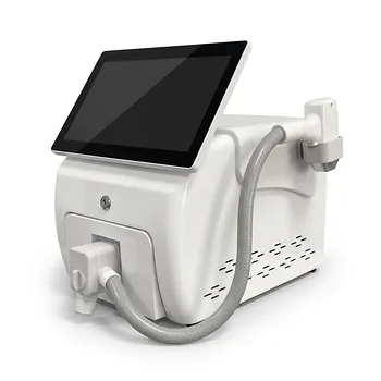
For dermal melanocytoses, safety depends more on controlled energy delivery and adequate healing time than on using the highest possible fluence. Technicians should perform a test spot, use conservative settings—especially for Fitzpatrick IV–VI skin—avoid pulse stacking and excessive overlap, and treat only to a uniform epidermal frosting endpoint. Strict cooling, sun protection, ocular protection, careful wound care, and treatment intervals of at least two months help reduce PIH, hypopigmentation, scarring, and other complications.
The safest protocol is test-guided and conservative: confirm the diagnosis, select wavelength and fluence for the lesion and skin type, treat to uniform frosting without excessive thermal injury, and wait long enough for inflammation and macrophage-mediated pigment clearance before repeating treatment.
Establish Whether Treatment Is Appropriate
Confirm the diagnosis and pigmentation pattern
Dermal melanocytoses include Nevus of Ota, Nevus of Ito, and Hori’s nevus. The diagnosis and depth of pigmentation should be confirmed by an appropriately qualified clinician before treatment, because facial hyperpigmentation may have other causes and may respond differently to laser therapy.
Hori’s nevus deserves particular caution because its melanocytes may be located near dermal blood vessels, increasing the likelihood of post-inflammatory hyperpigmentation.
Review medications and pigmentary risk
Obtain a complete medication and medical history before treatment. Drug-induced pigmentation, including conditions such as gold-related chrysiasis, can mimic or complicate pigmentary disorders and may not respond as expected to laser treatment.
Document the patient’s baseline skin tone, Fitzpatrick phototype, recent tanning, prior PIH, history of melasma, and previous laser reactions. Defer treatment when active inflammation, infection, sunburn, or uncontrolled tanning is present.
Prime and protect the skin when appropriate
For patients at elevated PIH risk, clinicians may consider a short pre-treatment course of a prescribed lightening agent such as hydroquinone or kojic acid, together with strict broad-spectrum UVA/UVB protection. Priming should be individualized and supervised rather than applied automatically to every patient.
Patients should avoid intentional tanning and use a broad-spectrum sunscreen, preferably with physical protection such as micronized zinc oxide. UV exposure before or after treatment can amplify inflammation and pigment production.
Select Conservative Laser Parameters
Use a test spot before full treatment
A test spot is essential, particularly for darker phototypes, facial lesions, Hori’s nevus, and patients with a history of PIH. Place it in a cosmetically inconspicuous area and observe both the immediate response and delayed pigmentary changes before treating the full lesion.
The test spot helps establish an appropriate fluence and reveals whether the patient develops excessive erythema, prolonged inflammation, hypopigmentation, or PIH.
Choose fluence by response, not by a fixed number
Use the lowest effective fluence that produces the intended clinical response. The supplementary material cites low settings around 2.5 J/cm² in some PIH-treatment contexts, but this should not be treated as a universal prescription for dermal melanocytoses; the correct value depends on wavelength, device, pulse duration, spot size, lesion depth, and skin phototype.
High fluence can injure surrounding tissue and stimulate melanocytes, particularly in Fitzpatrick IV–VI skin. Increase energy only cautiously and only when test-spot healing demonstrates that the tissue response is appropriate.
Target a uniform frosting endpoint
The desired immediate endpoint is uniform epidermal whitening or frosting across the treated area. Frosting results from rapid laser-induced tissue effects and generally fades within approximately 20 minutes.
Do not pursue excessive whitening, marked charring, widespread bleeding, or intense prolonged inflammation. These findings indicate unnecessary tissue injury and increase the risk of PIH, scarring, and permanent hypopigmentation.
Control beam distribution and coverage
A flat-top or top-hat beam profile and an appropriately large spot size can improve energy uniformity and reduce hot spots. The reference material gives spot sizes up to approximately 10 mm at 1064 nm as an example, but the correct size must match the device, lesion, anatomy, and manufacturer’s specifications.
Avoid pulse stacking and excessive spot overlap. Overlapping pulses can create localized thermal injury, dyschromia, and scarring, especially in darker skin.
Use a Controlled Treatment Technique
Apply cooling before and after treatment
Active cooling before treatment can reduce epidermal heating and discomfort. Apply cooling immediately afterward to limit burning, erythema, and inflammatory signaling.
Cooling should not obscure the endpoint or cause tissue injury from excessive cold. Use the device manufacturer’s recommended cooling method and monitor the skin continuously.
Provide appropriate analgesia
Q-switched laser treatment is painful, so topical anesthesia is commonly required. Apply it according to the product instructions, avoid excessive occlusion or prolonged exposure, and remove it completely before laser delivery.
Pain that is unexpectedly severe may indicate excessive fluence, inadequate cooling, or another technical problem and should prompt reassessment rather than simply increasing anesthesia.
Use wavelength-specific precautions
Select the wavelength according to the pigment’s depth, lesion characteristics, and the laser system’s validated indications. A 532 nm wavelength can cause transient purpura because of additional hemoglobin absorption, and this possibility should be discussed before treatment.
Near the orbit, use wavelength-appropriate ocular metal shields when indicated. The patient, operator, and all personnel in the room must use appropriate protection for the wavelength and treatment configuration; ordinary glasses are not an adequate substitute.
Maintain room and aerosol safety
Q-switched pulses can generate sparks, tissue splatter, and aerosolized debris. Keep flammable materials—including alcohol disinfectants, gauze, and oxygen tubing—out of the beam path.
Use appropriate masks, eye protection, and barriers or clear biologic dressings when indicated to contain debris. The room should follow applicable laser-safety, fire-safety, infection-control, and local regulatory requirements.
Schedule Sessions to Permit Healing
Wait at least two months between treatments
Dermal melanocytoses commonly require approximately four to eight sessions, but the number varies with lesion depth, size, skin type, and response.
Schedule treatments no more frequently than every two months. This interval allows inflammation to settle and gives macrophages time to clear fragmented pigment, while reducing the risk of cumulative thermal injury and permanent hypopigmentation.
Assess delayed reactions before retreating
Do not judge success solely by the immediate frosting response. Examine the patient after re-epithelialization and again after pigmentary changes have stabilized.
Retreatment should be postponed if erythema, crusting, PIH, hypopigmentation, or other inflammation persists. The next test spot or treatment should be adjusted based on the observed healing response.
Provide Structured Post-Treatment Care
Protect the epidermis during healing
Mild erythema, edema, stinging, and transient urticarial reactions may occur immediately. The treated area may then darken and develop a thin crust that usually flakes off over approximately 7–10 days.
Patients should cleanse gently with mild soap and water and apply petrolatum or another clinician-recommended protective ointment. They must not pick, scrub, or forcibly remove crusts.
Enforce strict UV avoidance
Use broad-spectrum UVA/UVB protection throughout healing and for several months afterward. Physical sunscreens containing micronized zinc oxide are specifically useful options, alongside hats, shade, and avoidance of deliberate sun exposure.
UV protection is one of the most important measures for preventing PIH and reducing recurrence or darkening of residual pigment.
Manage PIH cautiously
If PIH develops, clinicians may consider topical hydroquinone and strict photoprotection until the discoloration resolves. Treatment should begin only after assessing whether the skin has fully re-epithelialized and whether active inflammation or infection is present.
A short course of a prescribed anti-inflammatory combination may be used in selected cases, but prolonged or unsupervised topical steroid use must be avoided because it can cause skin atrophy and secondary hypopigmentation. Avoid adding irritating post-laser products, including tretinoin compounds, during the vulnerable healing period unless specifically directed by the treating clinician.
Counsel patients about expected effects
Explain that transient whitening, erythema, edema, crusting, and—particularly with 532 nm—purpura may occur. Set expectations that pigment reduction is gradual and commonly requires multiple sessions rather than one aggressive treatment.
Patients should contact the clinic for escalating pain, spreading redness, pus, blistering, delayed healing, significant scarring, or rapidly worsening pigmentation.
Understanding the Trade-offs
More energy does not necessarily mean better clearance
Higher fluence may produce stronger pigment disruption, but it also increases epidermal injury and inflammation. In darker skin, that inflammation can stimulate melanogenesis and produce PIH that is more noticeable or persistent than the original lesion.
A conservative, staged approach often provides better overall control than aggressive treatment followed by complications.
Frosting is useful but not a license to over-treat
Uniform frosting is the intended endpoint, but greater whitening is not a measure of greater efficacy. Excessive overlap, repeated pulses, or treatment of already inflamed skin can convert a controlled photothermal injury into unnecessary thermal damage.
Cooling and topical agents have limits
Cooling reduces discomfort and epidermal stress but cannot compensate for excessive fluence or poor technique. Similarly, bleaching agents may help manage pigmentary complications but do not eliminate the need for test spots, UV protection, and appropriate treatment intervals.
Hypopigmentation must also be prevented
Insufficient recovery time or repeated aggressive treatments can cause persistent or permanent hypopigmentation. This is why the two-month minimum interval and post-treatment assessment are central safety measures rather than scheduling preferences.
How to Apply This Safely in Practice
The following checklist should be adapted to the specific laser platform, lesion, skin type, and clinician’s governing protocols:
- If your primary focus is minimizing PIH: Use a conservative test-guided fluence, strict cooling, aggressive UVA/UVB protection, and avoid treatment while the skin is inflamed or recently tanned.
- If your primary focus is safe parameter selection: Base fluence, spot size, and overlap on the device, wavelength, skin phototype, and test-spot response—not on a fixed numerical setting.
- If your primary focus is preventing hypopigmentation: Avoid pulse stacking and excessive overlap, and separate sessions by at least two months to allow pigment clearance and tissue recovery.
- If your primary focus is periocular safety: Use wavelength-appropriate ocular protection, including metal shields when indicated, and restrict treatment to trained personnel following laser-safety procedures.
- If your primary focus is uncomplicated healing: Provide gentle cleansing, petrolatum or recommended protective ointment, no crust picking, and prompt review of abnormal pain, infection, blistering, or delayed healing.
A conservative, test-guided protocol with disciplined cooling, protection, follow-up, and spacing is the most reliable way to reduce complications while treating dermal melanocytoses.
Summary Table:
| Parameter | Recommended Approach | Rationale |
|---|---|---|
| Test spot | Always perform in inconspicuous area | Determines appropriate fluence and predicts PIH risk |
| Fluence | Lowest effective; use test spot response | High fluence increases PIH risk, especially in darker skin |
| Endpoint | Uniform epidermal frosting | Excessive whitening causes tissue injury |
| Spot size | Large enough for uniform coverage; flat-top beam | Reduces hot spots and uneven energy |
| Pulse stacking | Avoid | Causes localized thermal injury and dyschromia |
| Cooling | Active before and after | Reduces epidermal heating and inflammation |
| Treatment interval | At least 2 months | Allows healing and pigment clearance, reduces cumulative injury |
| Wavelength-specific | Use appropriate for depth; 532 nm may cause purpura | Matches laser to lesion characteristics |
| Ocular protection | Metal shields near orbit, eye protection for all | Prevents eye damage |
| Aerosol safety | Use masks, barriers; keep flammables away | Controls debris and fire risk |
At BELIS, we provide professional-grade Q-switched lasers and comprehensive training to help you achieve safe, effective treatments. Our advanced systems like the Nd:YAG and Pico lasers offer precise control for dermal melanocytoses. Contact us today to learn how our equipment and support can enhance your clinic's outcomes and patient safety.
Related Products
- Q Switch Nd Yag Laser Machine Tattoo Removal Nd Yag Machine
- Cryolipolysis Fat Freezing Cavitation Lipo Laser Machine
- Ultrasonic Cavitation Machine Lipo Laser Device
- Fractional CO2 Laser Machine for Skin Treatment
- Fractional CO2 Laser Machine for Skin Treatment
People Also Ask
- How does laser fluence influence pigment clearance vs. safety? Balancing Speed and Skin Integrity in Tattoo Removal
- What is the documented effectiveness of Q-switched Nd:YAG lasers for tattoo removal? Gold Standard Results
- What pigment compositions and particle properties should practitioners evaluate before operating Q-switched Nd:YAG lasers for tattoo removal? Evaluate pigment composition and particle size for safe, effective tattoo removal.
- How should wavelength and fluence settings be adjusted on Nd:YAG and Q-switched lasers when treating darker skin tones? Optimize Safety & Efficacy
- Which laser modalities and treatment schedules are recommended for clinical laser tattoo removal procedures? Q-Switched Nd:YAG and 6–12 Week Intervals Preferred for Safe, Effective Results