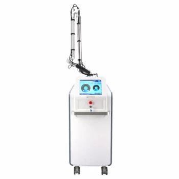
Excessive thermal injury is the central operational cause of scarring and demarcation lines during CO2 laser resurfacing. The highest-risk errors are excessive energy density, too many passes, overlapping pulses or scan patterns, inadequate cooling, and treating beyond the appropriate tissue endpoint. Sharp borders usually result from an abrupt change between fully treated and untreated skin, while scars and pigmentary changes reflect excessive or uneven heat accumulation.
The key prevention principle is controlled thermal delivery: use patient- and site-specific parameters, prevent pulse stacking and scan overlap, monitor tissue endpoints between passes, cool consistently, and reduce energy density at treatment borders through deliberate feathering.
How Operational Errors Produce Complications
Excessive energy density
High fluence or excessive impulse density can create a deep, thick zone of thermal necrosis. When injury extends too far into the dermis, the normal repair process may produce prolonged erythema, hypertrophic scarring, or keloid formation in susceptible patients.
The same excessive thermal load can also contribute to post-inflammatory hyperpigmentation or hypopigmentation, including marked “alabaster” hypopigmentation.
Too many consecutive passes
Repeated passes over the same region accumulate residual heat even when each individual pass appears acceptable. This is particularly hazardous when operators continue treating after the desired clinical endpoint has already been reached.
Pass counts should be limited according to the indication, treatment mode, anatomic site, and skin characteristics. Thin eyelid and neck skin generally require greater restraint than thicker cheek or perioral areas.
Overlapping pulses and scan patterns
Pulse overlap creates localized hot spots where energy density becomes higher than the nominal setting suggests. With automated scanners, imprecise alignment of adjacent patterns can similarly cause overlapping treatment fields and excessive focal heating.
In fractional treatment, overly high spot density can cause individual microscopic thermal zones to merge into a contiguous defect, effectively creating a full-field injury.
Stationary handpiece movement
Failing to keep the handpiece moving continuously can stack pulses in one location. This concentrates thermal energy and increases the risk of focal necrosis, persistent erythema, and scarring.
Steady movement and deliberate coverage are essential, particularly when using manual delivery.
Inadequate tissue cooling
Poor or inconsistent cooling allows heat to accumulate between passes. This can make otherwise reasonable settings unsafe, especially during high-energy treatment or in anatomically sensitive areas.
Cooling should be consistent with the device protocol and maintained throughout the procedure rather than applied only after excessive tissue reaction appears.
Why Demarcation Lines Develop
Abrupt treatment boundaries
A fully treated central field next to untreated surrounding skin can produce a stark visible transition. The greater the difference in energy delivery across that boundary, the more obvious the line may become during healing and repigmentation.
This is a field-design problem as much as a laser-setting problem.
Failure to feather the margins
Applying the same energy density to the center and edge of the treatment zone creates an abrupt endpoint. The peripheral border should instead be treated as a transition zone.
Practitioners can reduce fluence or energy density gradually toward the margins so the treated area blends into adjacent skin. This technique, known as feathering, reduces the visual contrast between treated and untreated tissue.
Uneven spacing or coverage
Gaps can leave untreated stripes, while excessive overlap can create darker, more inflamed lines or focal scars. Both problems result from inconsistent spacing and poor coordination between passes or scanner patterns.
Treatment patterns should be aligned precisely, with controlled spacing and no unintended overlap.
Controlling the Tissue Endpoint
Monitor the reaction between passes
Operators should assess tissue response after each pass rather than relying only on preset pass numbers. Vaporized or charred debris should be gently removed between passes with sterile saline-moistened gauze followed by dry gauze, allowing the underlying tissue to be evaluated accurately.
This improves visibility and reduces the risk of treating through debris that conceals the true endpoint.
Stop before excessive dermal injury
A faint chamois-yellow appearance is a warning that ablation has reached the reticular dermis and that further treatment substantially increases scarring risk. Treatment should stop when the appropriate clinical endpoint has been achieved, including disappearance of the target rhytid, visible desiccation, or the relevant tissue color change.
The correct endpoint depends on the device, technique, indication, and treatment area. A preset number of passes should never override direct tissue assessment.
Adjust for anatomy
The neck, chest, and eyelids are more vulnerable to scarring because they have thinner dermis and fewer adnexal structures than many facial areas. These regions generally require lower density, lower fluence, fewer passes, or a fractionated approach.
Parameters should therefore be selected by anatomic site rather than applied uniformly across the entire treatment area.
Preventing Uneven Thermal Delivery
Control pulse overlap
Operators should maintain the spacing specified for the device and treatment plan. Overlapping impulses too closely can increase local heat sufficiently to cause prolonged erythema, pigmentary change, or hypertrophic scarring.
The goal is predictable coverage, not maximal coverage at the highest possible density.
Align automated scanner patterns
Adjacent scan fields should meet precisely without overlapping. Scanner alignment errors can create linear areas of excess energy that later appear as prolonged erythema, pigment alteration, or scarring.
The operator should verify pattern placement throughout treatment, especially when moving between facial subunits or contour changes.
Remove debris between passes
Debris can obscure tissue reaction and interfere with accurate assessment of depth. Gentle cleansing between passes helps the operator identify whether additional treatment is justified.
Aggressive wiping should be avoided because mechanical trauma can add unnecessary injury to already compromised tissue.
Understanding the Trade-offs
More aggressive treatment is not always more effective
Higher energy, greater depth, and more passes may increase immediate resurfacing intensity, but they also increase cumulative thermal damage. Once treatment extends beyond the intended layer, additional energy can produce complications without providing proportional clinical benefit.
The practical objective is the lowest effective thermal dose for the specific indication and patient.
Uniform settings simplify execution but increase risk
Using one fluence, density, and pass count across the face may be operationally convenient. It ignores differences in skin thickness, vascularity, adnexal structures, and healing capacity.
Region-specific settings require more planning, but they provide better control over both scarring risk and visible transitions.
Fractionation reduces exposure but does not eliminate risk
Fractionated CO2 treatment can be useful for sensitive areas such as the neck because it limits the treated surface area. However, excessive pulse energy, depth, or spot density can still merge thermal zones and produce a near-contiguous injury.
Fractionation is a risk-management tool, not protection against overly aggressive parameters.
Postoperative factors can worsen an initial injury
Infection and patient scratching can convert a controlled injury into a more complicated wound-healing problem. Persistent erythema, pruritus, or early skin thickening should prompt early clinical assessment.
Any suspected hypertrophic scar or infection requires management according to the practitioner’s clinical protocol.
How to Apply This to Practice
Parameter selection should combine device settings, skin type, treatment indication, anatomic site, tissue response, and border design.
- If your primary focus is preventing hypertrophic scarring: Use conservative, site-specific energy and density settings, restrict pass counts, prevent pulse stacking and scan overlap, and stop at the appropriate tissue endpoint.
- If your primary focus is avoiding demarcation lines: Feather the peripheral treatment margins by gradually reducing energy density and maintain consistent spacing between treated and untreated zones.
- If your primary focus is treating thin or high-risk areas: Use fewer passes and lower-density or fractionated treatment on regions such as the eyelids, neck, and chest.
- If your primary focus is maintaining predictable coverage: Keep the handpiece moving, align scanner patterns precisely, control pulse overlap, and inspect tissue between passes after removing debris.
- If your primary focus is reducing pigmentary complications: Limit cumulative thermal injury, use appropriate cooling, and avoid excessive density or overlapping pulses, particularly in patients at higher risk of post-inflammatory hyperpigmentation.
Reliable CO2 resurfacing depends on controlling heat, respecting tissue endpoints, and designing treatment transitions deliberately rather than simply increasing treatment intensity.
Summary Table:
| Operational Error | Consequence | Prevention |
|---|---|---|
| Excessive energy density | Deep thermal necrosis, scarring, pigmentation | Use site-specific fluence, conservative settings |
| Too many passes | Accumulated heat, prolonged erythema, scarring | Limit passes, monitor endpoint between passes |
| Overlapping pulses/scan patterns | Focal hot spots, merges thermal zones | Adjust spacing, align scanner precisely |
| Stationary handpiece | Pulse stacking, focal necrosis | Keep handpiece moving continuously |
| Inadequate cooling | Tissue overheating, unsafe conditions | Follow device cooling protocol consistently |
| Abrupt treatment boundaries | Sharps demarcation lines | Feather margins: reduce fluence at edges |
| Uneven spacing/coverage | Untreated stripes, focal scarring | Control spacing, check coverage |
| Treating beyond tissue endpoint | Reticular dermis injury, scarring | Stop when chamois-yellow appears; remove debris |
| Not adjusting for anatomy | Higher risk in thin skin areas | Use lower density/fluence on neck, eyelids |
Ready to refine your laser technique and prevent complications? At BELIS, we offer advanced CO2 fractional laser systems designed for precise thermal control, along with comprehensive training and support. Our portfolio includes professional-grade aesthetic equipment trusted by clinics and premium salons worldwide. Contact us today to learn how our technology can help you achieve optimal results safely and efficiently. Get in touch with our experts →
Related Products
- Fractional CO2 Laser Machine for Skin Treatment
- Fractional CO2 Laser Machine for Skin Treatment
- Pico Laser Tattoo Removal Machine Picosure Picosecond Laser Machine
- Professional Face and Vaginal 7D HIFU System for HIFU Clinic Treatments
- Ultrasonic Cavitation Machine Lipo Laser Device
People Also Ask
- What is the technical principle behind CO2 Laser Fractional micro-perforations? Master Scar Revision Mechanics
- Why are professional laser systems, such as CO2 fractional or Erbium lasers, more effective for deep wrinkle reduction and dermal remodeling than topical anti-aging ingredients like retinoids, vitamin C, or peptides? Discover the targeted power of fractional lasers.
- What role does fractional CO2 laser equipment play in the treatment of SUI? Non-Surgical Stress Urinary Incontinence Care
- What is the rationale for a double-pass technique with fractional CO2 lasers? Maximize Deep Collagen Remodeling
- What is the purpose of manually extracting large cysts before CO2 fractional laser? Optimize Eyelid Milia En Plaque Care