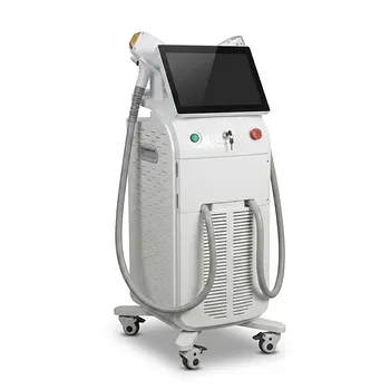
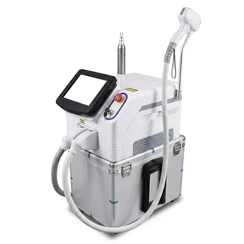
For small, sharply defined facial telangiectasia, clinicians generally favor a 532 nm KTP laser; for broader areas of diffuse erythema, PDL is often the better choice. The decision depends mainly on vessel size and depth, treatment area, and how much post-treatment purpura or downtime the patient will accept. KTP offers precise treatment with relatively little purpura, while PDL provides efficient coverage and strong results for diffuse superficial vascular redness.
The practical rule is simple: use KTP when the clinician needs precision vessel by vessel, and use PDL when the treatment objective is broader clearance of diffuse erythema or extensive superficial vascular change.
Start With the Vascular Pattern
Small, visible vessels favor KTP
KTP systems produce a 532 nm green wavelength, which is strongly absorbed by hemoglobin. Their small spot sizes allow the operator to trace and treat individual fine vessels directly.
This makes KTP particularly useful for discrete facial telangiectasia on the nose, cheeks, or other areas where precision matters. It is most appropriate when vessels are superficial, narrow, and visually well defined.
Diffuse erythema favors PDL
PDL is generally more efficient when the redness is spread over a large area rather than limited to isolated vessels. It is well suited to diffuse background erythema, including vascular redness associated with rosacea.
Larger spot sizes allow the clinician to treat broad regions more quickly and with fewer individual treatment passes.
Depth and vessel diameter influence the choice
Both systems are primarily used for superficial facial vascular lesions, but the clinician must still assess vessel depth and diameter. Very superficial, fine vessels are favorable targets for KTP, while PDL can address superficial telangiectasia across a wider field.
Deeper or larger vessels may require a different wavelength or pulse configuration, such as a long-pulsed 1064 nm Nd:YAG system, rather than simply increasing the settings on KTP or PDL.
Why KTP Is Chosen
It provides high targeting precision
KTP’s short penetration depth and small spot size can be advantageous when vessels are clearly visible and localized. The operator can follow the vessel path rather than treating the entire surrounding area.
This selective approach can reduce unnecessary exposure of adjacent skin and is particularly valuable for isolated nasal or facial vessels.
It can reduce visible downtime
KTP is commonly selected when minimizing purpura is a major priority. Its intended effect is controlled intravascular heating and coagulation rather than a pronounced mechanical disruption of the vessel wall.
Patients may still experience erythema, swelling, crusting, or pigmentary changes, but the risk of obvious bruise-like purpura is generally lower than with traditional PDL treatment.
It is less efficient for broad redness
The same precision that benefits individual vessels can become a limitation when the treatment area is extensive. Treating diffuse cheek erythema vessel by vessel may require more time and more passes than using a larger-area PDL approach.
KTP also has relatively shallow penetration, so it is not the best general solution for deeper or larger-caliber vessels.
Why PDL Is Chosen
It treats larger areas efficiently
PDL is designed to deliver energy across a treatment field, making it practical for diffuse erythema and widespread superficial telangiectasia. It is often preferred when the clinical problem is not a few isolated vessels but a broader vascular background.
This can make PDL more efficient for patients with rosacea-associated redness or extensive superficial facial vascularity.
It has a well-established vascular endpoint
The desired PDL endpoint is typically immediate vessel coagulation, darkening, or transient graying, depending on the device and treatment settings. The endpoint should be interpreted alongside the patient’s skin type and the level of epidermal protection available.
Settings must be individualized. Pulse duration, fluence, spot size, cooling, vessel diameter, and skin pigmentation all affect the balance between clearance and adverse effects.
Purpura is the main trade-off
Traditional PDL treatment can produce purpura, a bruise-like discoloration caused by vascular injury and shockwave effects. It may take days or, in some cases, up to approximately two weeks to fade.
Longer pulse durations and effective integrated epidermal cooling can reduce the likelihood or severity of purpura, although they do not eliminate it in every patient.
Match the Parameters to the Clinical Objective
KTP requires a conservative endpoint
For KTP treatment, clinicians generally begin with the lowest effective fluence and adjust according to the vessel response. A typical treatment goal is visible vasoconstriction or vessel clearing without excessive blanching or injury to the surrounding skin.
Reference ranges commonly include pulse durations around 10–30 ms, fluences approximately 10–20 J/cm² depending on spot size and device, and repetition rates in the range of roughly 3–8 Hz. These figures are not universal prescriptions because KTP platforms differ substantially in beam profile, spot size, cooling, and calibration.
PDL settings depend heavily on purpura tolerance
For superficial facial telangiectasia, published treatment approaches may use pulse widths around 6–10 ms, sometimes with pulse stacking, or longer durations around 20–40 ms. Fluence and spot size are adjusted to the vessel characteristics and desired endpoint.
Longer pulses may reduce purpura compared with more aggressive short-pulse treatment, but the clinician must ensure that the pulse duration remains appropriate for the target vessel diameter.
Cooling protects the epidermis
Active epidermal cooling is important with either platform, particularly when treating patients with higher melanin content or when using fluences sufficient to coagulate the vessel. Cooling helps protect the epidermis and may improve patient comfort.
It should not be used as a substitute for appropriate patient selection, conservative test spots, or careful endpoint monitoring.
Understand the Trade-offs
KTP: precision versus coverage
Advantages:
- Precise treatment of isolated, fine vessels.
- Generally lower risk of pronounced purpura.
- Useful when short visible downtime is important.
- Strong hemoglobin absorption at 532 nm.
Limitations:
- Less efficient for broad areas of diffuse erythema.
- Relatively shallow penetration.
- May be less suitable for deeper or larger vessels.
- Requires accurate vessel visualization and careful technique.
PDL: coverage versus downtime
Advantages:
- Efficient treatment of diffuse erythema.
- Useful for extensive superficial telangiectasia.
- Broad clinical experience in vascular lesion treatment.
- Flexible pulse durations and spot sizes.
Limitations:
- Purpura can be prominent, particularly with aggressive short-pulse treatment.
- Patients may require more downtime and concealment.
- Inadequate cooling or excessive fluence increases adverse-effect risk.
- It may be unnecessarily intensive for a few isolated fine vessels.
Do not choose by wavelength alone
A common mistake is to treat wavelength as the sole selection criterion. Clinical performance also depends on pulse duration, spot size, fluence, cooling, repetition rate, vessel diameter, skin type, and operator technique.
The same nominal wavelength can produce different outcomes on different systems. Device-specific protocols and test spots therefore remain essential.
Make the Choice Patient-Specific
Consider the patient’s downtime tolerance
A patient who needs minimal visible bruising may favor KTP when the target consists of a few superficial vessels. A patient willing to accept temporary purpura for more efficient treatment of diffuse redness may be a better PDL candidate.
The consultation should explicitly address the expected appearance after treatment, not just the anticipated clearance.
Assess whether the problem is focal or diffuse
The treatment map is often more informative than the diagnosis alone. A few linear vessels suggest KTP, while a broad field of redness suggests PDL.
Some patients have both patterns and may require staged or combined treatment, with each platform used for the lesion type it addresses most efficiently.
Account for skin pigmentation and vascular depth
Darker skin types require particular attention to epidermal melanin absorption and the risk of post-inflammatory hyperpigmentation. Conservative settings, effective cooling, and test spots become especially important.
If vessels appear deeper, larger, or resistant to superficial treatment, the clinician should reassess the wavelength rather than escalating superficial-device settings indefinitely.
Making the Right Choice for Your Goal
The best selection is based on the vascular pattern and the patient’s tolerance for downtime, not on the device label alone.
- If your primary focus is a few small, superficial, clearly visible vessels: Favor a 532 nm KTP system for precise vessel-by-vessel treatment with generally less purpura.
- If your primary focus is diffuse facial erythema or extensive superficial telangiectasia: Favor PDL for efficient coverage across a larger treatment area.
- If your primary focus is minimizing bruising and downtime: Consider KTP first for suitable focal vessels, or longer-pulse, well-cooled PDL settings when broader treatment is required.
- If your primary focus is deeper or larger-caliber vessels: Reassess whether a longer-wavelength platform, such as long-pulsed 1064 nm Nd:YAG, is more appropriate than either KTP or PDL.
The most reliable approach is to match the device and parameters to vessel depth, diameter, distribution, and the patient’s recovery priorities.
Summary Table:
| Feature | KTP (532 nm) | PDL |
|---|---|---|
| Best for | Small, well-defined vessels | Diffuse erythema, broad areas |
| Precision | High (small spot, vessel-specific) | Moderate (larger spot, field coverage) |
| Purpura risk | Lower | Higher (traditional) |
| Downtime | Generally less | May require more recovery time |
| Efficiency for broad areas | Lower | High |
| Typical pulse duration | 10–30 ms | 6–40 ms |
| Typical fluence | 10–20 J/cm² | Variable |
| Cooling | Important | Critical for epidermal protection |
Upgrade your clinic's vascular laser capabilities with BELIS' advanced diode, Alexandrite, and PDL systems – designed for precision and efficiency. Our professional-grade equipment, backed by expert support, ensures optimal outcomes for your patients. Discover how BELIS can elevate your practice – contact our specialists today for personalized recommendations and exclusive offers.
Related Products
- Cryolipolysis Fat Freezing Cavitation Lipo Laser Machine
- Cryolipolysis Fat Freezing Machine and Ultrasonic Cavitation Device
- Cryolipolysis Fat Freezing Machine Cavitation Lipo Laser Machine
- Tri Laser Diode Hair Removal Machine Professional Beauty Equipment
- Ultrasonic Cavitation Machine Lipo Laser Device
People Also Ask
- What are the main benefits of using a fat freezing machine? Achieve Permanent, Non-Surgical Body Sculpting
- Do fat freezing machines really work? Achieve Non-Invasive Body Contouring
- What are the potential side effects and risks associated with fat freezing? Safety Profile & Rare Risks Explained
- What is the treatment principle of cryolipolysis fat freezing? Science-Backed Fat Reduction and Body Contouring
- What is the recommended treatment course for different body parts using the cryolipolysis machine? Expert Fat Loss Tips