The key distinction is the reaction’s timeline and behavior after light exposure. A phototoxic reaction usually appears immediately or soon after exposure, peaks early, and then diminishes over 24–72 hours. A photoallergic reaction is typically delayed, becomes more intense over subsequent readings, and is more likely to produce itching and papulovesicular or eczematous changes.
Phototoxicity is generally an early, burning, sunburn-like reaction with a decrescendo pattern; photoallergy is a delayed, itchy, eczema-like reaction with a crescendo pattern. Skin diagnostic devices can document these patterns, but classification should rely on controlled phototesting, serial observations, clinical history, and—when necessary—medical evaluation.
Why the distinction matters before light-based procedures
Phototoxicity is a dose-related tissue injury
Phototoxic reactions occur when a light-sensitive substance absorbs optical radiation and directly increases tissue injury. They can resemble an exaggerated sunburn and may occur without prior sensitization.
Typical findings include erythema, warmth or burning, infiltration, and occasionally bullae in more severe reactions. The reaction generally appears immediately or after a short delay and then follows a decrescendo pattern, becoming less pronounced at later assessments.
Photoallergy is an immune-mediated response
Photoallergy involves an immune response to a substance that has been altered or activated by light. Prior sensitization may be required, so the reaction is not necessarily proportional to the amount of product or light received.
The onset is usually delayed. Findings more suggestive of photoallergy include pruritus, erythema, infiltration, papules, vesicles, or eczematous plaques, with a crescendo pattern that intensifies at 24, 48, or 72 hours.
The classification affects treatment planning
Both reactions can make a client unsuitable for immediate light or laser treatment. Misclassifying a delayed photoallergic reaction as mild transient erythema could expose the client to a procedure that worsens inflammation or increases the risk of pigmentary change and scarring.
How to use diagnostic devices and phototesting
Establish a controlled exposure and control field
A useful assessment compares small, defined areas exposed to controlled light doses with an unirradiated control field. The control helps determine whether the response is caused by the test substance, the light exposure, or an ordinary contact reaction.
A positive response in the unirradiated field immediately after patch removal suggests a contact reaction, rather than a purely light-dependent reaction. This distinction is important when evaluating sunscreens, topical medications, gels, or other products.
Record reactions at multiple time points
A single immediate reading is insufficient for reliably excluding photoallergy. Record the appearance and severity of each test area immediately or soon after exposure, then reassess at approximately 24, 48, and 72 hours, according to the clinic’s validated protocol.
Document:
- Onset: immediate, early, or delayed
- Peak: when the reaction is most severe
- Trajectory: decreasing or increasing over time
- Morphology: erythema, infiltration, papules, vesicles, bullae, erosions, or eczema
- Symptoms: burning, pain, or itching
- Distribution: limited to irradiated areas or also present in control areas
Serial documentation is more informative than the reaction’s initial intensity alone.
Apply a consistent reaction-grading system
Standardized grading improves communication and reduces subjective interpretation. A commonly used framework describes:
- Grade 1+: erythema
- Grade 2+: erythema with infiltration
- Grade 3+: erythema with papulovesicles
- Grade 4+: erythema with bullae or erosions
Phototoxic reactions often fall within the 1+ to 2+ range, although severity can vary with the photosensitizer and light dose. Photoallergic reactions are more suggestive when they are delayed, pruritic, and associated with 3+ or 4+ morphology, but grading must be interpreted alongside timing and clinical appearance.
Interpret device output as evidence, not a diagnosis
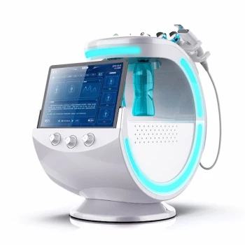
Imaging systems can help visualize erythema, surface changes, and the evolution of a reaction. They do not independently establish whether the mechanism is phototoxic or photoallergic.
The practitioner should combine device findings with the exposure protocol, product history, symptoms, control-field results, and serial progression. Suspected allergy or severe reactions warrant assessment by an appropriately qualified medical professional, often a dermatologist.
What to investigate during client screening
Review topical products and medications
Ask specifically about recent or current use of:
- Sunscreens and chemical UV filters
- Topical anti-inflammatory or antimicrobial agents
- Cosmetics and fragranced products
- Prescription and nonprescription medicines
- Supplements or systemic agents known to increase light sensitivity
Products containing substances such as PABA derivatives, oxybenzone, or camphor derivatives have been associated with contact or photoallergic reactions in susceptible individuals. The relevant exposure may be on the skin even when the client does not describe a history of “allergy.”
Ask about symptoms, not only visible redness
Burning or immediate discomfort supports a phototoxic, sunburn-like pattern. Prominent itching, especially when it develops later and persists or intensifies, raises greater concern for photoallergy.
Also ask about previous reactions to sunlight, tanning devices, laser treatments, sunscreens, or topical products. A history of chronic eczematous lesions on sun-exposed skin requires particular caution.
Consider baseline photosensitivity and skin condition
Standard skin typing does not fully predict an individual’s response to optical radiation. Barrier impairment, active dermatitis, chronic actinic dermatitis, and other photosensitivity disorders can increase risk independently of the client’s stated skin type.
If clinically indicated, a medical service may use tests such as minimal erythema dose assessment or formal photopatch testing to characterize sensitivity and identify a suspected photoactive substance.
Understanding the Trade-offs
Do not classify solely by reaction intensity
A severe early reaction is not automatically photoallergic, and a mild delayed reaction is not automatically harmless. Mechanism cannot be determined from grade alone.
The most reliable distinction comes from the combination of timing, trajectory, morphology, symptoms, and control-field findings.
Do not rely on one time point
A client may look clear immediately after testing and still develop a crescendo reaction over the next several days. Proceeding based only on an immediate device image is a major limitation of single-visit screening.
When delayed assessment cannot be completed, the practitioner should avoid treating a client with unexplained photosensitivity or a concerning product history rather than assuming the test is negative.
Do not confuse treatment erythema with diagnostic photosensitivity
Transient erythema can result from irritation, heat, pressure, or the procedure itself. Photoinduced reactions require interpretation in relation to the exposed and control areas, the applied substance, and the time course.
Atypical findings—including bullae, erosions, marked edema, systemic symptoms, or rapidly worsening inflammation—should not be managed as routine treatment effects.
Manage uncertainty conservatively
If the pattern is unclear, defer the light-based procedure, remove suspected photoactive products as clinically appropriate, and refer for medical evaluation. Selecting a lower fluence or adding cooling is not a substitute for resolving a suspected photosensitivity disorder.
How to Apply This to Your Screening Protocol
Use a documented, controlled, and serial approach rather than relying on device imaging or skin type alone.
- If your primary focus is rapid phototoxicity screening: Look for early burning, erythema, or infiltration that peaks early and follows a decrescendo pattern over 24–72 hours.
- If your primary focus is identifying photoallergy: Look for delayed, increasingly intense reactions with pruritus, papules, vesicles, or eczema-like changes during serial 24-, 48-, and 72-hour assessments.
- If your primary focus is separating light reactions from contact reactions: Compare irradiated and unirradiated fields, because a positive control field suggests contact sensitivity rather than a purely photoinduced response.
- If your primary focus is treatment safety: Defer treatment and seek appropriate medical assessment when reactions are severe, progressive, atypical, or inconsistent with a simple transient erythema.
- If your primary focus is individualized dosing: Establish baseline sensitivity through an appropriate medical phototesting protocol rather than relying exclusively on standardized skin typing.
A disciplined combination of controlled exposure, serial grading, morphology, symptoms, and clinical history is the safest way to distinguish phototoxicity from photoallergy before light-based aesthetic treatment.
Summary Table:
| Feature | Phototoxic | Photoallergic |
|---|---|---|
| Onset | Immediate or early | Delayed (24-72h) |
| Peak | Early | Late (crescendo) |
| Morphology | Sunburn-like (erythema) | Eczema-like (papules, vesicles) |
| Symptoms | Burning | Itching |
| Mechanism | Dose-related tissue injury | Immune-mediated |
| Sensitization required | No | Yes |
| Time course | Decrescendo | Crescendo |
Ensure your clients' safety with BELIS diagnostic devices. Our advanced imaging systems help you document and monitor skin reactions precisely. Contact us today to see how our devices enhance your screening protocols. Contact our team
Related Products
- Skin Tester Analysis Machine Analyser for Skin Testing
- Hydrafacial Machine with Facial Skin Analyzer Skin Tester
- Skin Tester Analysis Machine Analyser for Skin Testing
- Hydrofacial Machine with Facial Skin Analyzer and Skin Tester
- 4D 12D HIFU Machine Device for Skin Tightening
People Also Ask
- How does assessing pigment depth with advanced skin analysis devices impact laser parameter selection and patient outcomes in hyperpigmentation treatment? Optimize Laser Therapy Results
- What role do skin analysis and detection devices play in customized treatment? Enhance Precision and Patient Outcomes
- What is the function of skin analysis and diagnostic devices in medical aesthetic consultations? Scientific Data for Results
- How do skin testing and analysis devices assist in evaluating the effectiveness of fractional laser treatments?
- What role do skin testing and analysis devices play in managing patient expectations prior to laser and resurfacing procedures? Learn how objective data improves consultations and outcomes.