Infantile hemangiomas are dynamic vascular tumors that must be staged before laser treatment. Clinically, they progress from an early, often subtle precursor lesion through rapid proliferation, maturation, and eventual involution. Color-coded duplex sonography (CCDS) complements examination by showing whether vascularity is absent, emerging, highly active, or regressing. This distinction is essential because an actively proliferating lesion requires a different management strategy from residual telangiectasia or a noninvoluting vascular malformation.
The lesion’s biological phase, depth, and vascular flow pattern determine whether laser therapy is appropriate, which modality and parameters may be considered, and how complications such as ulceration or ineffective treatment can be avoided.
Why Staging Matters Before Treatment
Infantile Hemangiomas Are Not Static Lesions
Infantile hemangiomas usually develop after birth, enlarge rapidly during early infancy, and then undergo gradual involution over several years. Their appearance and internal blood flow can therefore change substantially over time.
A lesion that appears similar externally may be biologically active, stabilizing, or involuting. Treatment decisions based only on color or size can miss this distinction.
Treatment Must Follow Biological Behavior
During proliferation, endothelial activity and blood flow are high. The principal clinical priority may be controlling growth and preventing functional impairment, ulceration, or permanent distortion.
Systemic or topical beta-blocker therapy is the established medical approach for many clinically significant or high-risk infantile hemangiomas. Laser therapy may have a more limited, adjunctive role during active disease and a clearer role for selected superficial residual vessels after involution.
Staging Also Supports Differential Diagnosis
A true infantile hemangioma must be distinguished from a vascular malformation. Infantile hemangiomas typically show a period of rapid postnatal proliferation followed by involution, whereas vascular malformations are generally present at birth, grow with the child, and do not spontaneously regress.
This distinction is critical before laser treatment because the two conditions have different biology, prognosis, and treatment indications. Deep or diagnostically uncertain lesions may require CCDS and, in selected cases, MRI.
The Five Diagnostic Stages
1. Prodromal Phase
Clinically, the prodromal phase may appear as a small red or pale-white spot, localized telangiectasia, or a poorly defined swelling. The finding can be subtle and may not yet resemble a typical raised hemangioma.
CCDS may show a structureless, low-echo area without pathological vessels. At this point, the absence of demonstrable abnormal flow does not establish the final diagnosis by itself; clinical follow-up and reassessment may be necessary.
2. Initial Phase
The initial phase is characterized by loss of normal skin architecture, increasing thickness, and developing induration. The lesion begins to become more clinically recognizable as a vascular tumor.
On CCDS, the center may remain hypoechoic, while hypervascularity begins at the margins. This peripheral vascular development can indicate that the lesion is entering an active growth period.
3. Proliferation Phase
Clinically, the lesion commonly becomes bright red, more infiltrated, and increasingly elevated or extensive. Subcutaneous growth may accompany the superficial component, producing a mixed or deeper lesion.
CCDS typically demonstrates marked intratumoral hyperperfusion, dense central vessel networks, and feeding vessels with arterial flow profiles. These findings are consistent with active vascular proliferation rather than a stable residual lesion.
4. Maturation Phase
During maturation, growth slows and the lesion may become pale, violaceous, or livid. Central ulceration may occur, particularly in vulnerable locations or lesions exposed to friction, moisture, or pressure.
Sonographically, central vessel density declines and the central component becomes increasingly hyperechoic. The reduction in flow suggests that the lesion is transitioning away from its most active proliferative state.
5. Regression Phase
Regression is associated with hypopigmentation, wrinkled or redundant skin, and residual palpable induration. The lesion may become flatter while leaving telangiectasia, fibrofatty tissue, atrophy, or textural changes.
CCDS shows circumscribed hyperechoic areas with loss of the typical tumor vascularity. A residual red surface does not necessarily indicate ongoing tumor proliferation; it may represent superficial telangiectasia or post-involutionary change.
How Clinical and Sonographic Findings Work Together
Clinical Examination Defines the Visible Phenotype
Examination assesses timing, color, elevation, surface changes, ulceration, distribution, depth clues, and effects on nearby structures. The history is particularly important because the sequence of postnatal appearance and growth often distinguishes an infantile hemangioma from a congenital malformation.
Anatomical site also affects urgency. Lesions near the eye, airway, nose, lips, or other functionally important structures may require specialist assessment even when their surface area is limited.
CCDS Assesses Internal Activity
CCDS adds information that cannot be obtained reliably from inspection. It helps characterize lesion depth, vascular density, perfusion, feeding vessels, and the transition from active flow to involution.
For a proliferating hemangioma, hyperperfusion and spontaneous internal flow support active disease. By contrast, vascular malformations may show different architecture, including heterogeneous tissue or multilocular vascular spaces, depending on the malformation type.
The Combination Reduces Diagnostic Error
No single color, ultrasound appearance, or treatment response should be interpreted in isolation. Clinical history, examination, CCDS, and MRI when indicated provide a more reliable diagnostic picture.
This is especially important for deep subcutaneous lesions, mixed superficial and deep lesions, and lesions that were historically labeled “cavernous hemangiomas” but may actually represent venous malformations.
Matching Stage to Laser Considerations
Active Proliferation Requires Caution
High-flow, highly vascular lesions are not automatically suitable for direct aesthetic laser treatment. In a rapidly growing or high-risk hemangioma, delaying effective medical management in favor of laser treatment can allow avoidable progression.
If laser is considered, the decision should be made by an appropriately trained specialist within a broader treatment plan. Lesion depth, location, ulceration status, skin integrity, and concurrent pharmacological treatment all influence risk.
Superficial Residual Vessels May Be More Suitable
After substantial involution, persistent superficial telangiectasia or erythematous residual vessels may be considered for selected vascular laser treatments. At this stage, the target is often residual cutaneous vascularity rather than the proliferating tumor itself.
The expected benefit is therefore different: treatment may improve color and visible vessels but cannot necessarily correct fibrofatty residuum, redundant skin, scarring, or deeper structural changes.
Depth Influences Modality Selection
A superficial lesion and a lesion with substantial subcutaneous involvement do not present the same optical or thermal target. Wavelength, spot size, pulse duration, fluence, cooling, and treatment interval must be selected according to the target vessel and tissue depth.
A deeper-penetrating system may be considered in selected deeper vascular targets, but greater penetration also increases the risk of unintended thermal injury. Device selection must follow diagnosis and anatomy rather than the device name alone.
Understanding the Trade-offs
Laser Is Not a Universal First-Line Treatment
The supplementary material correctly emphasizes that laser can be useful in selected circumstances, but it overstates the idea that early laser routinely “inhibits” hemangioma proliferation or accelerates complete involution. Current management commonly prioritizes observation for low-risk lesions and beta-blocker therapy for lesions requiring active treatment.
Laser may be appropriate for ulceration in specific settings, selected superficial disease, or residual telangiectasia. The indication should be individualized rather than inferred from the presence of visible redness.
Treating the Wrong Lesion Can Be Ineffective
A vascular malformation will not follow the natural proliferative and involutionary course of an infantile hemangioma. Applying a superficial laser to a deep venous malformation may produce limited benefit while exposing the child to pain, pigmentary change, scarring, or thermal injury.
Likewise, treating post-involutionary fibrofatty tissue as though it were active vascular tumor is unlikely to achieve the intended result.
Ulceration Changes the Risk Profile
Ulceration is not simply a cosmetic feature. It indicates compromised skin integrity and may involve pain, bleeding, infection risk, and delayed healing.
Laser parameters that might be considered for intact superficial vessels may be inappropriate over ulcerated or fragile tissue. Treatment planning should account for wound care and the possibility that medical management or specialist intervention takes priority.
Ultrasound Findings Have Limits
CCDS is valuable, but interpretation depends on equipment settings, lesion depth, operator experience, and the quality of the examination. Low detectable flow does not always prove complete biological inactivity.
When findings and clinical behavior disagree, repeat imaging, specialist review, or MRI may be warranted rather than immediate laser treatment.
Making the Right Choice for Your Goal
The practical decision should be based on diagnosis, developmental stage, risk category, depth, and treatment objective.
- If your primary focus is diagnostic accuracy: Combine the postnatal growth history and physical examination with CCDS, and use MRI when a deep or ambiguous lesion cannot be adequately characterized.
- If your primary focus is controlling active growth: Prioritize specialist risk assessment and evidence-based medical management; do not assume that aesthetic laser is a substitute for beta-blocker therapy.
- If your primary focus is treating residual redness: Confirm that the lesion is largely involuted and that the remaining target is superficial telangiectasia rather than active deep tumor or fibrofatty residuum.
- If your primary focus is minimizing treatment complications: Use the documented vascularity, depth, skin condition, and anatomical site to guide modality, pulse duration, fluence, cooling, and treatment timing.
Accurate staging turns laser treatment from a device-driven procedure into a diagnosis-driven clinical decision.
Summary Table:
| Stage | Clinical Features | Sonographic Findings | Laser Consideration |
|---|---|---|---|
| Prodromal | Small red/pale spot, telangiectasia | Hypoechoic, no pathological vessels | Observe, re-evaluate |
| Initial | Induration, loss of architecture | Hypervascular margins | Monitor closely |
| Proliferation | Bright red, elevated | Marked hyperperfusion | Caution; medical therapy |
| Maturation | Pale/violaceous, slower growth | Decreased central vessels | Evaluate for residual vessels |
| Regression | Flattening, fibrofatty tissue | Hyperechoic, avascular | Selective for telangiectasia |
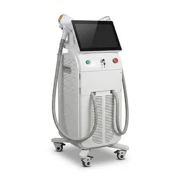
Are you a clinic or aesthetic professional seeking to refine hemangioma laser protocols? BELIS offers advanced vascular laser systems (Nd:YAG, Pico, IPL) with precision features suited for staged treatment. Our team provides clinical training and device selection guidance. Contact us today to optimize your outcomes and expand your aesthetic service capabilities.
Related Products
- 9D 7D HIFU Vaginal RF Lifting Treatment
- IPL SHR+Radio frecuency machine
- Professional Face and Vaginal 7D HIFU System for HIFU Clinic Treatments
- 4D Vaginal HIFU and Face HIFU System
- IPL SHR Hair Removal Machine for Permanent Hair Removal
People Also Ask
- Is HIFU treatment considered safe and what are the potential risks? A Guide to Safe Non-Invasive Skin Tightening
- Is a HIFU treatment painful? Discover Comfort Levels and Results for Non-Surgical Lifting
- How does HIFU treatment differ from other skin tightening procedures? Compare Non-Invasive Lifting vs. Surgery
- What are the primary benefits of HIFU treatment? Reveal a Youthful V-Line and Firm Skin Non-Invasively
- Why are HIFU systems equipped with multiple treatment handpieces? Precision Depth Control for Superior Skin Results