Ablative Er:YAG and pulsed CO2 lasers can provide definitive treatment for facial eccrine hidrocystomas by vaporizing the cyst wall rather than merely draining its contents. This approach reduces recurrence compared with needle aspiration or simple incision and drainage. Because these lesions often occur on or near the eyelids, treatment requires wavelength-appropriate eye protection and careful control of laser exposure around the ocular surface.
Ablative laser treatment works by removing the cyst wall, which is the structure responsible for recurrence. Its effectiveness depends on complete, controlled ablation and strict ocular, plume, fire, and wound-care safety protocols.
How Ablative Lasers Treat Eccrine Hidrocystomas
The Cyst Wall Is the Treatment Target
An eccrine hidrocystoma is a benign cystic lesion associated with sweat ducts. Aspiration removes fluid temporarily, but leaving the cyst wall behind allows the lesion to refill.
Er:YAG and pulsed CO2 lasers ablate or vaporize the cyst wall under direct visualization. Complete removal of this lining is the key reason laser treatment can produce a more durable result than drainage alone.
Er:YAG and Pulsed CO2 Have Different Tissue Effects
Er:YAG laser energy is strongly absorbed by water, allowing precise superficial vaporization with relatively limited residual thermal damage. This can be useful when the lesion is small and located close to delicate eyelid structures.
Pulsed CO2 lasers also use water absorption to ablate tissue, but generally produce more thermal coagulation around the treatment zone. That thermal effect can assist hemostasis but requires careful control near the eyelid and ocular surface.
Treatment Requires Direct Visual Control
The clinician should identify the lesion boundaries and ablate the cyst wall in a controlled manner. Treatment should stop once the wall has been adequately removed, avoiding unnecessary injury to surrounding skin.
Because eyelid lesions can be close to the visual axis, this procedure should be performed by a clinician trained in both laser tissue effects and periorbital anatomy.
Why Laser Ablation May Reduce Recurrence
Drainage Does Not Eliminate the Lining
Needle aspiration and simple incision can decompress a hidrocystoma, but they do not reliably remove the secretory cyst wall. The lesion may therefore recur after an initially successful result.
Laser ablation addresses the underlying structure rather than only its contents. This is the central therapeutic advantage of the technique.
Precision Matters More Than Aggressiveness
The goal is complete treatment of the cyst wall with the least possible collateral damage. Excessive energy, repeated passes, or unnecessarily deep ablation can increase inflammation, delayed healing, pigmentary changes, and scarring.
The appropriate laser, pulse settings, spot size, and number of passes must be selected according to lesion depth, location, and the clinician’s equipment-specific protocol.
Required Safety Protocols
Protect the Patient’s Eyes
Patient eye protection is mandatory for lesions near the eyelids or visual axis. Appropriate metal external or corneal shields should be selected for the laser wavelength and positioned correctly before laser emission begins.
Plastic shields should not be used where the laser could strike them, because they may melt or fail to provide adequate protection. The shield must be placed without trapping debris or causing pressure injury to the cornea.
Protect Clinical Personnel
All operating personnel should wear protective eyewear appropriate to the laser wavelength. Eyewear must remain in place whenever the laser is armed or capable of emitting energy.
The treatment room should have controlled access, clear laser warning signage, and a designated laser safety process. Only trained personnel should operate or assist with the system.
Control Laser Plume
Ablation creates a plume containing vaporized tissue and potentially hazardous biological material. A high-efficiency smoke evacuator should be used with its capture opening positioned as close as practical to the treatment site, ideally within approximately 1 cm.
Laser masks or respirators appropriate to institutional policy should supplement, not replace, active plume evacuation. Staff should also follow local infection-control and occupational-safety requirements.
The strongest evidence for infectious plume concerns involves procedures on viral lesions; the level of risk varies by tissue and pathogen, and claims of proven HIV transmission through laser plume should not be overstated.
Prevent Fire and Equipment Hazards
The room should have immediate access to water and an appropriate fire extinguisher. Flammable materials, alcohol-based residues, dry gauze, and oxygen-enriched conditions should be managed carefully.
If an airway device is used, any exposed endotracheal tube should be protected according to the anesthesia and laser-safety protocol, including appropriate wet saline gauze where indicated. The laser should be placed in standby whenever it is not actively being used.
Prepare the Skin Correctly
Topical or local anesthesia may be required because ablation removes superficial tissue. Any topical anesthetic must be completely removed before laser emission.
Because ablative laser wavelengths are highly absorbed by water, the treatment surface should be dried according to the device protocol. Cooling, including directed forced-cold air when appropriate, can improve comfort and help limit unwanted thermal injury.
Postoperative Care and Monitoring
Support Re-epithelialization
For a small localized lesion, the clinician may recommend a bland wound-healing ointment or another suitable non-irritating emollient until re-epithelialization is complete. The aim is to reduce crust formation and maintain a controlled healing environment.
Routine topical antibiotics are not automatically required and can cause contact dermatitis. The chosen product should follow the treating clinician’s protocol and the patient’s allergy history.
Avoid Excessive Occlusion
Thick or excessive occlusive application can contribute to folliculitis or acneiform eruptions. Patients should apply only the amount directed and should not repeatedly layer products over the area without instruction.
Petrolatum-based products are flammable in the presence of an open flame. Patients using them should avoid smoking, candles, and other ignition sources during recovery.
Monitor for Complications
Patients should receive instructions about expected redness, swelling, crusting, and healing time. They should promptly report increasing pain, spreading redness, purulent drainage, significant swelling, visual symptoms, or delayed healing.
Periorbital swelling and ocular symptoms deserve particular attention because the treatment site is close to the eye. Any suspected corneal injury or visual change requires urgent ophthalmic assessment.
Understanding the Trade-offs
Recurrence Is Reduced, Not Impossible
Complete cyst-wall ablation can substantially reduce recurrence, but no technique guarantees that recurrence cannot occur. Incomplete treatment, multifocal disease, or an incorrect diagnosis may lead to persistence or regrowth.
Histologic assessment may be appropriate when the diagnosis is uncertain, the lesion is atypical, or the clinical appearance does not clearly match a benign hidrocystoma.
Cosmetic Injury Remains Possible
Ablative treatment can cause prolonged erythema, post-inflammatory hyperpigmentation or hypopigmentation, infection, delayed healing, and scarring. These risks are more consequential on thin eyelid skin and in patients prone to abnormal pigmentation or poor wound healing.
The expected cosmetic benefit must therefore be weighed against the lesion’s size, symptoms, recurrence history, and proximity to the eye.
Laser Is Not Appropriate for Every Facial Cyst
A lesion that is infected, solid, atypical, deeply situated, or diagnostically uncertain may require another evaluation or treatment strategy. Ablative laser should not be selected solely because a lesion is cosmetically visible.
Diagnosis and treatment planning should precede laser use, particularly for lesions near the eyelid margin or lacrimal structures.
How to Apply This to the Treatment Decision
The appropriate approach depends on the lesion’s diagnosis, location, depth, recurrence pattern, and cosmetic importance.
- If your primary focus is durable treatment: Favor complete, controlled ablation of the cyst wall over fluid aspiration alone, provided the diagnosis and anatomy are suitable.
- If your primary focus is ocular safety: Require correctly placed metal eye shields, wavelength-specific staff eyewear, controlled laser access, and a periorbital protocol before treatment begins.
- If your primary focus is minimizing tissue injury: Use the lowest effective, device-appropriate treatment parameters and stop once the cyst wall has been adequately treated.
- If your primary focus is uncomplicated healing: Follow a prescribed bland wound-care regimen, avoid excessive occlusion, and monitor closely for infection, scarring, or visual symptoms.
- If your primary focus is occupational safety: Use active plume evacuation near the treatment point, appropriate respiratory protection, fire controls, and formal laser-safety procedures.
Ablative laser treatment is most effective when definitive cyst-wall removal is combined with disciplined periorbital, plume, fire, and postoperative safety practices.
Summary Table:
| Aspect | Er:YAG Laser | Pulsed CO2 Laser |
|---|---|---|
| Tissue Effect | Precise superficial vaporization with minimal thermal damage | Vaporization with more thermal coagulation, aiding hemostasis |
| Best For | Small lesions near delicate eyelid structures | Lesions where hemostasis is preferred |
| Safety Consideration | Requires eye protection and careful control near ocular surface | Similar ocular risks; more thermal spread requires careful control |
| Recurrence | Complete ablation of cyst wall reduces recurrence | Complete ablation of cyst wall helps prevent refilling |
| Common Adverse Effects | Erythema, pigment changes, scarring | Similar, plus potentially more thermal injury |
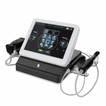
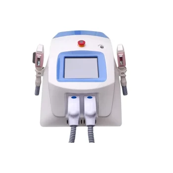
Enhance your clinic's treatment capabilities with advanced laser systems from BELIS. Our professional-grade Er:YAG and CO2 fractional lasers are designed for precise, safe, and effective procedures. Learn how our technology can benefit your practice and patients — contact us today to discuss your needs and discover our full range of aesthetic equipment.
Related Products
- Fractional CO2 Laser Machine for Skin Treatment
- Fractional CO2 Laser Machine for Skin Treatment
- Clinic Use IPL and SHR Hair Removal Machine with Nd Yag Laser Tattoo Removal
- Clinic Use IPL SHR ND YAG Laser Hair Removal RF Skin Tightening Machine
- Professional Face and Vaginal 7D HIFU System for HIFU Clinic Treatments
People Also Ask
- What is the technical principle behind CO2 Laser Fractional micro-perforations? Master Scar Revision Mechanics
- What parameters and treatment intervals are advised when applying fractional CO2 laser technology to delicate periorbital skin laxity? Discover safe protocols for eyelid rejuvenation.
- How do energy-based modalities complement injectable neurotoxins in décolleté rejuvenation? Explore synergistic benefits.
- What is the primary function of a high-precision fractional CO2 laser system for GSM? Restore Vaginal Health Naturally
- Why do fractional CO2 laser parameters need to be differentiated? Master Keloid vs. Hypertrophic Scar Treatment