The precision of picosecond laser therapy for Xanthelasma Palpebrarum depends entirely on the accuracy of clinical documentation. Standardized imaging equipment is necessary because it ensures consistency in shooting distance, lighting, and angles, providing the only objective basis for quantifying lesion clearance and adjusting laser parameters. Without this level of control, clinicians cannot accurately measure progress or maintain the high safety standards required for delicate periorbital treatments.
Standardized imaging transforms subjective visual observation into objective, scientific data. This precision allows for the quantification of lesion clearance rates and provides the necessary evidence for the dynamic optimization of laser energy parameters throughout the treatment cycle.
Establishing a Scientific Baseline for Evaluation
Eliminating Environmental Variables
The primary role of standardized equipment is to fix shooting distance, lighting conditions, and angles. By removing these variables, clinicians ensure that changes observed in the skin are due to the treatment itself rather than differences in how the photograph was taken.
Enabling Quantitative Analysis
Standardized images provide a scientific basis for quantifying lesion clearance rates, often graded at 25%, 50%, or 75% intervals. This high-resolution documentation allows for the use of the Visual Analog Scale (VAS) to assess progress objectively rather than relying on clinician memory.
Precision Archival Documentation
Precise records serve as a core technical tool for quality control within medical institutions. These archives provide a verifiable trail of treatment efficacy that can be used for internal audits and managing patient expectations.
Optimizing Clinical Decision-Making
Dynamic Adjustment of Laser Parameters
Standardized imaging allows practitioners to see how the tissue responded to previous settings. This data is essential for the dynamic adjustment of laser energy parameters, ensuring that subsequent sessions are tailored to the patient's specific healing trajectory.
Identifying Clinical Endpoints
High-resolution systems help clinicians accurately determine clinical endpoints, such as erythema response, pigment darkening, or crusting. Recognizing these signs early allows the practitioner to refine the energy levels and filter selections for the next treatment phase.
Monitoring Tissue Remodeling
By comparing pre- and post-treatment images, professionals can track the skin remodeling progress in the delicate eyelid area. This objective visual baseline is critical for verifying that the laser is effectively targeting the xanthelasma without causing unnecessary trauma to the surrounding dermis.
Understanding the Trade-offs and Pitfalls
The Risk of Subjective Bias
Without standardized equipment, clinical evaluation is prone to subjective bias. Variations in ambient light can make a lesion appear lighter or darker than it actually is, leading to incorrect energy settings that could result in either undertreatment or tissue damage.
Technical Limitations of Consumer Hardware
Relying on standard digital cameras or smartphones often fails to capture the subtle nuances of pigment depth and skin texture. This lack of detail can obscure early signs of adverse effects, making it difficult to intervene before a complication becomes permanent.
Equipment Cost vs. Clinical Safety
While standardized imaging systems require a higher initial investment, the cost of mismanaged parameters—such as unintended depigmentation—is significantly higher. Precise documentation is a vital risk-management tool that protects both the patient’s health and the provider’s reputation.
Applying Standards to Your Practice
To maximize the efficacy of picosecond laser treatments for Xanthelasma Palpebrarum, imaging protocols should be integrated into every stage of the clinical workflow.
- If your primary focus is treatment safety: Use standardized imaging to monitor for early signs of adverse reactions like depigmentation or unintended hair loss in the brow area.
- If your primary focus is clinical efficacy: Rely on high-resolution comparisons to calculate precise clearance grades and adjust energy fluences for subsequent sessions.
- If your primary focus is patient satisfaction: Utilize the objective visual baseline to provide patients with clear evidence of their progress, which helps manage expectations throughout the multi-session process.
Standardized imaging is the bridge between clinical intuition and scientific precision, ensuring every laser pulse is backed by objective data.
Summary Table:
| Feature | Standardized Imaging | Subjective/Manual Evaluation |
|---|---|---|
| Consistency | Fixed lighting, distance, and angles | Variable environmental conditions |
| Data Quality | High-resolution, quantitative data | Qualitative, memory-based assessments |
| Parameter Optimization | Dynamic adjustment based on healing | Guesswork or fixed protocols |
| Clinical Endpoints | Precisely identified (erythema/crusting) | Hard to distinguish subtle changes |
| Risk Management | Early detection of adverse effects | High risk of unintended depigmentation |
Elevate Your Clinical Precision with BELIS
To achieve the highest safety standards in delicate periorbital treatments like Xanthelasma removal, your clinic needs more than just a laser—it needs a professional-grade ecosystem. BELIS specializes in providing exclusive, high-performance medical aesthetic equipment for clinics and premium salons.
Our advanced portfolio empowers practitioners with:
- Precision Lasers: Pico, Nd:YAG, Alexandrite, CO2 Fractional, and Diode Hair Removal systems.
- Diagnostic & Care: Advanced skin testers for standardized imaging and Hydrafacial systems.
- Advanced Modalities: HIFU, Microneedle RF, and body sculpting (EMSlim, Cryolipolysis).
Ready to upgrade your treatment outcomes? Contact us today to discover how BELIS equipment can bring scientific precision and superior ROI to your practice.
References
- Fakültesi Su, Liuqing Chen. Optimizing Laser Therapy: Efficacy and Safety of Picosecond 1,064 nm Nd:YAG Laser in Xanthelasma Palpebrarum Treatment. DOI: 10.1155/dth/6693871
This article is also based on technical information from Belislaser Knowledge Base .
Related Products
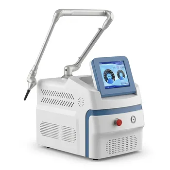
- Pico Picosecond Laser Machine for Tattoo Removal Picosure Pico Laser
- Pico Laser Tattoo Removal Machine Picosure Picosecond Laser Machine
People Also Ask
- What is the physical mechanism behind the high-decibel popping sound of picosecond lasers? Ink Shattering Physics
- What key role does the Diffractive Optical Element (DOE) play in picosecond laser skin reconstruction? Expert Analysis
- What role does picosecond laser equipment play in tattoo removal? Faster Results & Advanced Precision
- What is the mechanism of Picosecond or Q-switched lasers for melasma? Advanced Photoacoustic Pigment Removal
- What are the core advantages of high-performance picosecond laser equipment? Superior tattoo removal for your clinic.