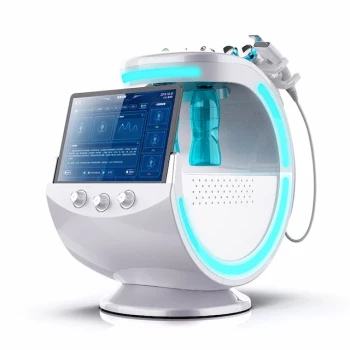
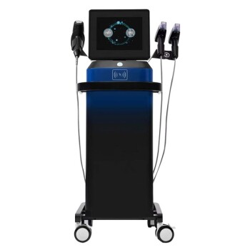
Professional skin testing devices turn photoaging assessment from a primarily visual judgment into a measurable clinical evaluation. During consultations, multispectral imaging and polarized-light analysis can help document telangiectasia, pigment distribution, roughness, and early structural changes that may be difficult to see under ordinary lighting. This information supports treatment selection, establishes a baseline, and allows clinicians to track change across a rejuvenation program.
Photoaging is multidimensional, so reliable assessment requires more than judging visible wrinkles. Professional analyzers provide objective images and quantitative indicators that help clinicians understand the location, severity, and treatment response of vascular, pigmentary, and textural abnormalities.
Why Objective Photoaging Assessment Matters
Photoaging affects more than the skin surface
Photoaging can involve uneven melanin distribution in the epidermis, dermal collagen and elastin degradation, superficial vascular dilation, roughness, and reduced translucency.
Some of these changes are visible during a standard examination, while others are subsurface or too subtle to evaluate consistently by eye. A device helps reveal patterns that may otherwise be underestimated.
Visual examination is useful but limited
Clinical observation remains essential because it incorporates color, morphology, symptoms, history, and physical examination. However, it can be affected by lighting, camera settings, observer experience, and the patient’s immediate skin condition.
Device-based imaging adds repeatable documentation and gives the consultation a more consistent reference point.
How Devices Evaluate Telangiectasia
Mapping vascular dilation
Cross-polarized, multispectral, or other specialized imaging approaches can emphasize superficial vascular structures and areas of vascular dilation. This may help identify the distribution and relative prominence of telangiectasia across the face.
The resulting images can show whether vascular changes are localized or widespread, and whether they coexist with pigmentation or generalized photodamage.
Supporting vascular treatment decisions
A vascular map can contribute to decisions about whether light-based or laser therapy should be considered and which areas require particular caution. It may also help clinicians distinguish a predominantly vascular presentation from one in which redness is mainly associated with irritation or inflammation.
The device does not replace clinical diagnosis. It provides additional evidence that must be interpreted alongside history, examination, and the patient’s treatment goals.
Establishing a vascular baseline
Baseline imaging makes it easier to compare telangiectasia before and after treatment. This is particularly valuable when improvement is gradual or when the patient’s perception is influenced by temporary redness, swelling, or changes in lighting.
Consistent photography and standardized imaging conditions are important for making these comparisons meaningful.
How Devices Evaluate Pigmentation
Measuring pigment distribution
Professional systems can visualize and quantify visible and subsurface pigmentation patterns using tools such as multispectral imaging, polarized light, or Wood’s lamp examination.
They may help document the distribution of hyperpigmented macules, uneven melanin deposition, and deeper pigment patterns that are not fully apparent under standard illumination.
Supporting differential assessment
Imaging can contribute to the clinical distinction between conditions such as solar lentigines, melasma, and post-inflammatory hyperpigmentation by showing differences in distribution, morphology, and apparent depth.
This information is supportive rather than definitive. A device image alone should not be treated as a diagnosis, particularly when lesions are atypical, changing, or clinically concerning.
Improving treatment planning
Quantified pigmentation data can help clinicians decide whether a patient may be better suited to a pigment-targeting laser, light-based treatment, chemical peel, topical program, or a staged combination approach.
It also helps identify cases in which aggressive treatment could increase the risk of post-inflammatory hyperpigmentation, especially when the patient has a higher risk profile or an unstable pigment disorder.
How Devices Evaluate Textural Changes
Documenting roughness and microrelief
Photoaging-related texture includes superficial roughness, fine lines, wrinkle depth, enlarged irregularities, and loss of smooth light reflection. High-resolution imaging can document these changes more consistently than unaided inspection.
Some systems generate surface maps or numerical indicators related to wrinkle depth and texture roughness. These measures provide a structured way to describe changes over time.
Detecting early structural trends
Textural deterioration may develop before deep wrinkles become obvious. Device imaging can help identify early fine-line patterns, uneven surface relief, or broader changes in skin microstructure.
This is useful when the consultation is focused on prevention or early intervention rather than correction of advanced photoaging.
Connecting texture to treatment selection
Texture findings can help clinicians determine whether the main treatment objective is superficial resurfacing, collagen remodeling, hydration and barrier support, or a combination of approaches.
The data should guide treatment intensity, but it should not be used in isolation. Skin sensitivity, healing history, medications, phototype, and contraindications remain equally important.
How Testing Devices Improve the Consultation
Creating a reproducible baseline
Pre-treatment imaging records the patient’s starting condition, including pigment distribution, vascular features, wrinkle patterns, and surface irregularity.
This baseline is especially important before laser resurfacing, light-based therapy, chemical peeling, or multi-stage rejuvenation protocols.
Setting realistic expectations
Patients often focus on the most visible concern, such as a dark spot or wrinkle, while overlooking associated vascular or textural changes. Showing objective images can clarify that photoaging is a combination of findings rather than a single defect.
It also helps clinicians explain which changes are likely to improve, which may require multiple sessions, and which may be managed rather than completely eliminated.
Tracking treatment response
Follow-up imaging can quantify or visually document changes that are difficult to judge from memory. This may include reduced pigment contrast, less visible vascular dilation, shallower wrinkle patterns, or improved surface uniformity.
For valid comparisons, the clinic should standardize lighting, camera distance, facial expression, skin preparation, and the interval between assessments.
Supporting personalized protocols
The combined assessment can help clinicians tailor the treatment plan to the patient’s dominant photoaging parameters. A patient with primarily vascular changes may need a different strategy from one with deep pigmentation, rough texture, or mixed findings.
This supports more deliberate selection of treatment modality, treatment depth, energy level, and sequencing.
Understanding the Trade-offs
Quantification does not equal diagnosis
A numerical score can create an impression of precision without proving the underlying diagnosis. Device outputs depend on the instrument, software, lighting conditions, calibration, and the way the data are interpreted.
Clinical judgment remains necessary, particularly for differentiating benign photoaging from lesions that require medical evaluation.
Results are not automatically comparable
Images taken with different devices, different settings, or inconsistent preparation may produce misleading comparisons. Recent tanning, makeup, topical products, erythema, hydration changes, and treatment-related inflammation can all affect the result.
A clinic should use a consistent imaging protocol and interpret trends rather than relying on a single score.
Algorithms have boundaries
Automated systems may identify patterns, but they may not account fully for skin tone, active inflammation, mixed pigmentation, or unusual lesion morphology. Results should therefore be treated as decision support, not as an independent treatment prescription.
More data can complicate the consultation
Presenting every available metric may overwhelm patients or distract from the clinically important findings. The most useful consultation translates device data into a small number of actionable conclusions.
How to Apply This to Clinical Practice
A professional skin analysis system is most valuable when it is integrated into the consultation workflow rather than used as a stand-alone sales or scoring tool.
- If your primary focus is telangiectasia: Use standardized vascular imaging to map redness and visible vessels, then interpret the findings with a clinical examination before selecting or recommending vascular treatment.
- If your primary focus is pigmentation: Use multispectral, polarized, or Wood’s lamp imaging to document pigment distribution and support differentiation among common pigmentary patterns.
- If your primary focus is textural change: Use high-resolution imaging or surface analysis to record roughness, fine lines, and wrinkle depth, then match treatment intensity to the patient’s skin condition and recovery capacity.
- If your primary focus is treatment planning: Combine vascular, pigmentary, and textural findings with medical history, phototype, and contraindication screening to create a staged, individualized protocol.
- If your primary focus is proving treatment effectiveness: Capture a standardized baseline and repeat imaging under comparable conditions so that improvement can be assessed objectively over time.
Used appropriately, professional skin testing devices make photoaging consultations more consistent, more transparent, and more clinically actionable.
Summary Table:
| Parameter | Device Capability | Clinical Utility |
|---|---|---|
| Telangiectasia | Map vascular dilation via cross-polarized imaging | Support vascular treatment decisions and baseline establishment |
| Pigmentation | Quantify melanin distribution using multispectral imaging | Differentiate pigmentary patterns and guide treatment planning |
| Textural Changes | Document surface roughness and wrinkle depth | Identify early structural trends and tailor resurfacing intensity |
Elevate your consultation precision with BELIS professional skin testing devices. Our advanced imaging solutions provide objective data for telangiectasia, pigmentation, and texture analysis, enabling tailored treatment plans for your clinic. Join leading clinics and premium salons that trust BELIS for cutting-edge aesthetic technology. Contact us today to discuss how our devices can enhance your practice and patient outcomes. Get in touch to schedule a demo.
Related Products
- Skin Tester Analysis Machine Analyser for Skin Testing
- Skin Tester Analysis Machine Analyser for Skin Testing
- Hydrofacial Machine with Facial Skin Analyzer and Skin Tester
- Hydrafacial Machine with Facial Skin Analyzer Skin Tester
- 4D 12D HIFU Machine Device for Skin Tightening
People Also Ask
- What role do skin analysis and detection devices play in customized treatment? Enhance Precision and Patient Outcomes
- How do skin testing and analysis devices assist in evaluating the effectiveness of fractional laser treatments?
- What is the function of skin analysis and diagnostic devices in medical aesthetic consultations? Scientific Data for Results
- What role do skin testing and analysis devices play in managing patient expectations prior to laser and resurfacing procedures? Learn how objective data improves consultations and outcomes.
- How does assessing pigment depth with advanced skin analysis devices impact laser parameter selection and patient outcomes in hyperpigmentation treatment? Optimize Laser Therapy Results