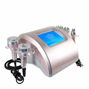
UVA-assisted skin testers help clinicians make vitiligo more visible and clinically distinguishable. Devices operating at 320–400 nm, commonly called Wood’s illumination, accentuate complete epidermal depigmentation and can reveal subtle lesions that are difficult to see under ordinary lighting. They support—not replace—clinical examination by helping practitioners assess lesion borders, pigment loss, and patterns associated with vitiligo versus other hypopigmentation disorders.
The central value of UVA illumination is contrast: it makes fully depigmented vitiligo patches appear more sharply defined while helping clinicians recognize conditions that produce incomplete, irregular, or secondary pigment reduction.
How UVA Illumination Improves Vitiligo Assessment
It Increases Contrast Between Normal and Affected Skin
Normal skin contains melanin that absorbs and scatters light differently from depigmented skin. Under UVA illumination, vitiligo lesions typically appear bright, sharply contrasted, or chalky white, making their margins easier to identify.
This is particularly useful when lesions are subtle, located on sun-protected areas, or present on darker skin tones.
It Highlights Complete Melanin Loss
Vitiligo is characterized by the loss or destruction of functional melanocytes, producing depigmentation rather than merely reduced pigmentation. UVA examination helps the clinician determine whether a pale area represents near-complete pigment absence or only partial pigment reduction.
This distinction is clinically important because post-inflammatory hypopigmentation and similar disorders may retain pigment, even when they look white under ordinary room lighting.
It Can Reveal Fluorescence Associated With Vitiligo Lesions
Abnormal accumulation of biopterin metabolites in vitiligo skin can produce characteristic fluorescence under UVA exposure. This optical response can further increase the contrast between affected and unaffected skin.
The fluorescence pattern should be treated as a supporting clinical sign, not as an independent confirmation of vitiligo.
What Clinicians Examine During the Test
Border Definition and Lesion Shape
Vitiligo commonly presents as well-demarcated macules or patches with clear borders. UVA illumination can make these borders more apparent and help define the true extent of a lesion before treatment or documentation.
Irregular, mottled, or poorly defined areas may point toward another cause, although lesion appearance must always be interpreted in the wider clinical context.
Distribution and Symmetry
Practitioners should assess whether lesions follow patterns commonly associated with vitiligo, including acral, periorificial, and joint-extensor locations. Symmetrical distribution can support the clinical impression of vitiligo.
Distribution alone is not diagnostic, because several inflammatory, infectious, chemical, and post-traumatic conditions can produce overlapping patterns.
Residual Pigment and Perifollicular Repigmentation
A UVA examination may help reveal areas of residual pigment within a pale lesion or around hair follicles. These findings can assist with assessing the degree and pattern of pigment loss and may also provide a useful baseline for monitoring repigmentation.
The device should therefore be used not only for initial observation but also for standardized follow-up images under consistent conditions.
Differentiating Vitiligo From Common Mimics
Post-Inflammatory Hypopigmentation
Hypopigmentation following eczema, psoriasis, lupus, or another inflammatory process often appears irregular or mottled, with residual pigment and sometimes adjacent hyperpigmentation. UVA illumination can make this incomplete pigment loss easier to distinguish from the more uniform depigmentation typical of vitiligo.
A history of preceding inflammation, scaling, itching, or treatment is essential to the interpretation.
Pityriasis Versicolor
Pityriasis versicolor may produce lighter or darker patches with subtle scale, commonly on the trunk and upper limbs. UVA examination can improve visual contrast, but it does not by itself establish the diagnosis.
When infection is suspected, clinicians should combine the examination with appropriate testing, such as direct microscopy or other validated methods, rather than treating the lesion as vitiligo solely because it appears pale.
Chemical Leukoderma
Chemical leukoderma can resemble vitiligo, particularly when exposure affects the hands, face, or other contact areas. A careful occupational, cosmetic, and medication history is necessary because the key diagnostic clue may be ongoing or prior exposure to phenolic or related chemicals, not the optical appearance alone.
UVA illumination helps map the depigmented areas, but identifying and stopping the causative exposure remains central to management.
Other Infectious or Inflammatory Disorders
Leprosy and other conditions can produce hypopigmented lesions that overlap visually with vitiligo. Sensory changes, inflammation, scale, systemic symptoms, exposure history, and lesion distribution may provide more diagnostic information than fluorescence alone.
If the findings are atypical, clinicians may need laboratory testing, dermoscopy, or biopsy.
Why Accurate Differentiation Matters
It Prevents Inappropriate Light-Based Treatment
Not every pale lesion should receive phototherapy, repigmentation treatment, or aesthetic intervention. Treating an infection, active inflammation, or chemical injury as vitiligo can delay the correct management and potentially worsen the underlying condition.
A confirmed or well-supported diagnosis allows the practitioner to select a safer and more targeted protocol.
It Establishes a Reliable Treatment Baseline
High-resolution imaging under consistent UVA conditions can document lesion borders, distribution, and apparent pigment intensity. This creates a reference for monitoring progression, stabilization, or repigmentation over time.
Baseline documentation is especially valuable when lesions are subtle or when treatment response will be evaluated over multiple visits.
It Supports Assessment Across Different Skin Tones
Visual examination under white light can underestimate pigment loss, particularly in darker complexions or areas with limited contrast. UVA illumination provides an additional optical view that can improve lesion detection and boundary mapping.
It should complement, rather than replace, examination under normal lighting.
Understanding the Trade-offs
UVA Findings Are Supportive, Not Definitive
Wood’s illumination cannot reliably distinguish every cause of hypopigmentation. A bright or sharply contrasted lesion may support vitiligo, but diagnosis still depends on history, morphology, distribution, symptoms, and—when indicated—additional testing.
The safest interpretation is clinical diagnosis supported by UVA imaging, not automated diagnosis based on fluorescence alone.
Device Quality and Technique Affect Results
Results depend on the device’s wavelength range, optical output, image quality, room darkness, working distance, and skin preparation. Ambient light, cosmetics, topical products, and inconsistent positioning can reduce contrast or create misleading artifacts.
Clinics should use validated equipment and consistent examination protocols.
Fluorescence Can Be Misinterpreted
Different biological materials can fluoresce under ultraviolet illumination. Porphyrin-related red fluorescence, for example, is not the same finding as the enhanced white contrast used to assess depigmentation and should not be interpreted as evidence of vitiligo.
Practitioners must distinguish between pigment contrast and fluorescence from other tissue or microbial products.
Safety and Scope Must Be Controlled
Diagnostic UVA exposure should be brief and performed according to the equipment manufacturer’s instructions and appropriate clinical safety procedures. The device is primarily an assessment aid; it should not be treated as a substitute for a dermatologist or for confirmatory investigations when the presentation is uncertain.
How to Apply This to Clinical Practice
Use UVA-assisted skin testing as part of a structured differential-diagnosis workflow:
- If your primary focus is detecting subtle vitiligo: Examine the skin under controlled UVA illumination to identify sharply demarcated, fully depigmented areas that may be inconspicuous under white light.
- If your primary focus is distinguishing depigmentation from hypopigmentation: Compare lesion contrast, residual pigment, border regularity, and clinical distribution rather than relying on brightness alone.
- If your primary focus is excluding mimics: Combine UVA findings with medical history, scale or inflammation assessment, sensory testing, exposure review, and targeted laboratory tests when infection or chemical leukoderma is possible.
- If your primary focus is monitoring treatment: Capture standardized baseline and follow-up images using the same device settings, room conditions, and anatomical views.
- If your primary focus is treatment safety: Do not begin phototherapy or aesthetic procedures solely because a lesion appears bright under UVA; refer atypical, progressive, symptomatic, or uncertain cases for specialist evaluation.
Used correctly, UVA illumination turns subtle pigment differences into clinically useful evidence while keeping final diagnosis grounded in comprehensive patient assessment.
Summary Table:
| Feature | Vitiligo Under UVA | Other Hypopigmentation Disorders |
|---|---|---|
| Pigment Loss | Complete, sharply defined | Partial, irregular, or mottled |
| Border Definition | Well-demarcated | Often ill-defined |
| Fluorescence | May show biopterin-related fluorescence | Usually absent |
| Residual Pigment | Minimal or perifollicular | Often present |
| Distribution | Acral, periorificial, symmetrical | Varies by cause |
| Key Diagnostic Clue | Depigmentation pattern | History, inflammation, exposure, or infection |
Ready to enhance your diagnostic accuracy with professional UVA skin testers? BELIS offers advanced imaging devices designed for clinics and premium salons, helping you precisely assess vitiligo and other skin conditions. Our portfolio includes state-of-the-art skin testers and a full range of medical aesthetic equipment. Contact us today to learn how our solutions can elevate your practice and patient outcomes — Get in touch with our experts.
Related Products
- Skin Tester Analysis Machine Analyser for Skin Testing
- Skin Tester Analysis Machine Analyser for Skin Testing
- Hydrofacial Machine with Facial Skin Analyzer and Skin Tester
- Hydrafacial Machine with Facial Skin Analyzer Skin Tester
- Ultrasonic Cavitation Radiofrecuency Machine for Body Slimming
People Also Ask
- What role do skin testing and analysis devices play in managing patient expectations prior to laser and resurfacing procedures? Learn how objective data improves consultations and outcomes.
- How do skin testing and analysis devices assist in evaluating the effectiveness of fractional laser treatments?
- What role do skin analysis and detection devices play in customized treatment? Enhance Precision and Patient Outcomes
- How does assessing pigment depth with advanced skin analysis devices impact laser parameter selection and patient outcomes in hyperpigmentation treatment? Optimize Laser Therapy Results
- How can diagnostic evaluation frameworks and skin analysis devices be used to guide parameter selection for facial resurfacing procedures? Unlock Personalized Treatment Planning