For oral mucosal vascular lesions, safety depends on conservative energy delivery, reliable cooling, and airway planning. A 755 nm Alexandrite system may be used for selected deep or bulky lesions, but clinicians should not transfer cutaneous protocols directly to the tongue, lips, or airway-adjacent mucosa. A representative reference setting is 40 J/cm², 3 ms, and a 12 mm spot size with dynamic cooling; final parameters must be individualized to the lesion, device, tissue thickness, and treatment response.
Treat oral lesions conservatively and in a controlled setting: use validated dynamic cooling, avoid pulse stacking and overlap, monitor the endpoint closely, and prepare for significant edema whenever the lesion is near the airway.
Why Oral Mucosa Requires a More Conservative Protocol
Deep penetration increases the risk of hidden thermal injury
The 755 nm wavelength penetrates more deeply than shorter-wavelength vascular lasers. This can be useful for thick, violaceous, or submucosal lesions, but the absence of dramatic immediate surface change does not indicate that insufficient energy was delivered.
Thermal injury may develop below the visible surface. Clinicians should therefore avoid escalating fluence simply because the lesion does not immediately appear fully treated.
Edema can become an airway emergency
Aggressive photothermal treatment of the tongue, floor of mouth, or other airway-adjacent sites can cause severe localized edema. In extreme cases, swelling may compromise or occlude the airway.
Treatment should occur only where the practitioner can assess and manage airway risk. Lesion location, size, depth, baseline airway status, and the anticipated need for anesthesia or airway support should be evaluated before treatment.
Confirm the diagnosis and treatment suitability
A vascular-appearing oral lesion should be clinically assessed before laser treatment. The practitioner should consider lesion depth, compressibility, bleeding behavior, prior growth, and whether tissue diagnosis or imaging is needed.
Laser treatment should not substitute for diagnostic evaluation when the diagnosis is uncertain or when features are atypical.
Parameter Guidelines for 755 nm Treatment
Use conservative starting parameters
For a deep mucosal lesion, the primary reference describes 40 J/cm², 3 ms, and a 12 mm spot size with dynamic cooling as an example protocol. This should be treated as a reference point, not a universal prescription.
The appropriate setting depends on lesion caliber, depth, color, tissue thickness, and the specific laser’s pulse profile and cooling system. A test area or limited initial treatment is prudent when the device and lesion combination is unfamiliar.
Match spot size and fluence
The general relationship is important:
- Larger spots, approximately 12–15 mm: generally paired with lower fluences, around 25–35 J/cm².
- Smaller spots, approximately 6–8 mm: may be paired with higher fluences, around 50–80 J/cm², when clinically justified.
- Short pulses, such as 3 ms: may be used for selected smaller or resistant lesions, but they deliver energy rapidly and require particularly careful monitoring.
- Longer pulses, approximately 3–20 ms: may be selected according to lesion depth and vessel caliber.
These ranges come largely from vascular skin and lip applications. They should not be applied automatically to intraoral mucosa, where tissue response and airway consequences differ.
Use lesion-specific caution
For lip venous lakes, published reference guidance describes a 6 mm spot, approximately 3 ms pulse, and 60–80 J/cm², with an endpoint of slight immediate shrinkage or subtle bluing.
For a bulky or deep lesion on the tongue, the lower-fluence, larger-spot example—40 J/cm², 3 ms, 12 mm—is more consistent with a cautious initial approach. The treating clinician must confirm that these settings are supported by the device manufacturer and appropriate for the specific mucosal site.
Cooling and Delivery Technique
Treat dynamic cooling as essential
Dynamic cooling, such as cryogen spray cooling, is identified as essential for protecting superficial, non-target tissue while treating deeper vascular components. It reduces the risk of epidermal or superficial mucosal thermal injury when therapeutic fluences are used.
For intraoral use, the cooling method must be compatible with the device, treatment site, airway plan, and manufacturer’s instructions. A system validated for external skin should not automatically be assumed safe or practical inside the mouth.
Prevent pulse stacking and excessive overlap
Do not repeatedly fire pulses over the same point or create dense overlap in a small area. The 755 nm wavelength can produce cumulative bulk heating even when each individual pulse appears acceptable.
Use controlled, contiguous coverage only when appropriate, and document the treated area so that subsequent sessions do not unintentionally repeat excessive exposure.
Do not chase a dramatic visual endpoint
The immediate endpoint may be subtle, such as transient gray discoloration, slight shrinkage, or subtle bluing. A deeper lesion may later develop persistent deep purpura as vascular injury evolves.
The absence of immediate whitening or dramatic vessel disappearance is not a reason to increase energy aggressively. Stop or reassess if the tissue response suggests excessive heating, blistering, marked swelling, or unexpected pain.
Periprocedural Safety Protocol
Prepare for airway and swelling risk
Before treatment, establish a specific plan for managing post-treatment edema. This is particularly important for lesions involving the tongue, base of tongue, floor of mouth, soft palate, or tissues close to the airway.
High-risk cases may require treatment in a facility with appropriate airway expertise and rescue capability rather than a routine office environment.
Use appropriate laser safety controls
Apply standard Class 4 laser controls, including:
- Correct wavelength-specific eye protection for everyone in the controlled area.
- A designated laser-controlled zone with warning signage.
- Restricted access during laser activation.
- Appropriate nonreflective instruments and surfaces.
- Smoke or plume control when tissue vaporization or thermal plume is possible.
- A trained assistant who can monitor the patient, equipment, and airway concerns.
For intraoral treatment, eye protection must protect against the 755 nm wavelength without interfering with the operator’s ability to visualize the lesion.
Protect adjacent structures
Use appropriate barriers and careful positioning to protect teeth, restorative materials, uninvolved mucosa, and adjacent skin. Avoid directing energy toward reflective instruments or unintended tissue.
If a cooling spray is used, confirm that it cannot pool, obscure the field, interfere with airway management, or create a risk of aspiration.
Obtain informed consent
Consent should address the possibility of pain, swelling, bruising or purpura, ulceration, delayed healing, scarring, pigmentary change, incomplete clearance, recurrence, and the possibility of staged treatment.
For airway-adjacent lesions, consent should specifically include the risk of clinically significant edema and the potential need for urgent airway management.
Understanding the Trade-offs
Higher fluence is not automatically better
Higher fluence may improve treatment of resistant or deeper vascular components, but it also increases the risk of epidermal or mucosal injury, bulk heating, edema, scarring, and pigmentary complications.
The safest strategy is usually the lowest effective exposure that produces an appropriate delayed vascular endpoint.
755 nm is not ideal for every vascular lesion
Small, superficial, light-red vessels may respond poorly because hemoglobin absorption at 755 nm is lower than at shorter visible wavelengths. Deeper or thicker, violaceous lesions are more logical targets for this wavelength.
A different wavelength or modality may be more appropriate when the lesion is very superficial, when diagnostic uncertainty remains, or when the required energy would create unacceptable mucosal risk.
Darker skin increases pigmentary risk
Although oral mucosa differs from facial or cutaneous skin, adjacent pigmented skin and the patient’s overall phototype remain relevant to thermal and pigmentary risk. In cutaneous applications, Fitzpatrick skin types I–III are generally more favorable, while types IV–VI have greater risk of dyspigmentation.
Cooling, conservative parameters, and avoidance of unnecessary treatment of surrounding pigmented tissue are especially important.
A single session may be insufficient
Deep lesions often require staged treatment rather than one aggressive session. Attempting complete clearance in one treatment increases the chance of excessive edema and collateral injury.
The interval and number of sessions should be determined by the clinical response, healing, residual lesion burden, and the safety of re-treatment—not by a fixed schedule.
How to Apply This to Your Clinical Protocol
Use the following framework rather than treating the parameter ranges as a substitute for clinical judgment:
- If your primary focus is airway safety: Treat tongue and airway-adjacent lesions conservatively in an appropriately equipped setting, with a documented edema and airway-management plan.
- If your primary focus is parameter selection: Begin with a device-validated, conservative protocol; a reference example is 40 J/cm², 3 ms, and 12 mm with dynamic cooling, followed by response-based adjustment.
- If your primary focus is thermal protection: Use validated dynamic cooling, avoid pulse stacking and excessive overlap, and do not escalate energy merely because the immediate endpoint is subtle.
- If your primary focus is treatment efficacy: Reserve 755 nm primarily for suitable deep, bulky, or violaceous vascular lesions and consider staged treatment rather than aggressive single-session delivery.
- If your primary focus is diagnostic safety: Confirm that the lesion is appropriate for laser treatment and obtain imaging, specialist assessment, or biopsy when the diagnosis or depth is uncertain.
A disciplined, conservative protocol—supported by cooling, endpoint monitoring, and airway preparedness—provides the safest path to treating oral mucosal vascular lesions with a 755 nm Alexandrite laser.
Summary Table:
| Parameter | Guideline |
|---|---|
| Fluence | 40 J/cm² (reference for deep lesions) |
| Pulse duration | 3 ms (reference) |
| Spot size | 12 mm (reference) |
| Cooling | Essential, dynamic cooling |
| Endpoint | Subtle shrinkage, gray discoloration |
| Session | Staged, avoid aggressive single treatment |
Ensure safe and effective treatment with BELIS's advanced 755 nm Alexandrite laser systems, designed for clinical excellence. Contact our experts to learn how our devices can enhance your practice's vascular lesion management.
Related Products
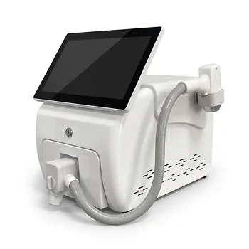
- Tri Laser Diode Hair Removal Machine Professional Beauty Equipment
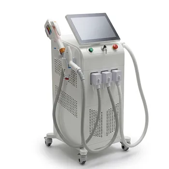
- 808nm Diode Laser Hair Removal Machine 755+808+1064nm Mixed Wavelength Professional Equipment
- 9D 7D HIFU Vaginal RF Lifting Treatment
- Professional Face and Vaginal 7D HIFU System for HIFU Clinic Treatments
People Also Ask
- What ocular safety protocols are required when operating diode or alexandrite hair removal lasers near the periorbital area? Essential eye protection and shielding for safe treatment.
- How do 800/810 nm diode hair removal lasers compare to 1064 nm long-pulsed Nd:YAG lasers? Discover the Best for Your Patients
- How do 800–810 nm Diode hair removal lasers compare to 1064 nm Long-Pulsed Nd:YAG lasers in terms of hair reduction efficacy and patient comfort? Discover Key Differences for Optimal Hair Removal
- What role does Thermal Relaxation Time (TRT) play when setting pulse duration parameters on diode laser hair removal and vascular laser equipment? Optimize treatments for better outcomes.
- Why is active skin cooling critical during high-energy aesthetic laser procedures such as diode laser hair removal? Avoid Burns, Pigment Changes, and Scarring