High-precision digital imaging systems are technically necessary because they replace subjective visual estimation with standardized, objective data. By strictly controlling variables such as lighting, distance, and angles, these systems capture subtle morphological changes that verify the actual efficacy of scar repair treatments. This rigorous approach eliminates human bias, allowing clinicians to grade outcomes based on measurable evidence like depression depth and color consistency rather than opinion.
The core value of high-precision imaging lies in its ability to generate a scientific baseline for evaluation. It transforms scar assessment from a qualitative art into a quantitative science, serving as the definitive method for validating the clinical performance of medical aesthetic equipment.
The Engineering of Objectivity
Eliminating Environmental Variables
The primary technical failure in manual scar assessment is environmental inconsistency. Shadows and ambient light can skew the perception of scar depth and texture.
High-precision systems mitigate this by using standardized lighting environments, often employing ring flashes or LED arrays. This ensures that every image—whether pre-treatment or post-treatment—is captured under identical illumination, eliminating interference from external light sources.
Consistency in Focal Geometry
Beyond lighting, the physical geometry of the capture must remain constant. These systems utilize fixed focal lengths and specific shooting angles to maintain a consistent field of view.
This standardization ensures that changes observed in the image reflect actual biological changes in the tissue, rather than differences in camera distance or perspective.
From Visuals to Quantitative Data
Micron-Level Surface Analysis
While the human eye can see general improvement, it cannot quantify it. Advanced digital systems, particularly those with 3D analysis capabilities, provide micron-level measurements of skin topography.
By mapping the skin's surface, these systems generate quantitative data regarding roughness and scar volume. This allows for the precise tracking of depression depth recovery throughout the treatment cycle.
Visualizing Structural Changes
To make fine details accessible, high-precision systems often employ sophisticated software to generate color-coded mappings of the skin.
These maps translate complex topographic data into visual formats that highlight fine structural changes and pigment distribution. This visualization is critical for performing histopathological correlation analysis, linking visual outcomes to underlying tissue changes.
Understanding the Operational Trade-offs
The Requirement for Strict Protocol
The technical advantage of these systems relies entirely on protocol adherence. Because the systems are designed to detect micron-level changes, even slight deviations in the setup—such as a shift in patient positioning or angle—can introduce artifacts into the data.
Complexity vs. Speed
Unlike a simple photograph, utilizing a high-precision system with macro lenses and 3D analysis software adds a layer of complexity to the clinical workflow.
Obtaining high-definition details of fine lines and texture requires proper calibration and focus. The trade-off for scientific accuracy is a requirement for operational discipline; these systems are not "point-and-shoot" solutions but precision instruments that demand correct usage to yield valid data.
Making the Right Choice for Your Goal
To maximize the value of digital imaging in scar evaluation, align your technology use with your primary objective:
- If your primary focus is Clinical Research: Prioritize systems that offer quantitative, micron-level data to prove efficacy and eliminate inter-observer variability in your studies.
- If your primary focus is Patient Communication: Utilize systems that feature color-coded mapping and high-definition texture visualization to clearly demonstrate progress that may be subtle to the naked eye.
True clinical excellence in scar repair requires measuring the invisible to prove the visible.
Summary Table:
| Feature | Manual Visual Assessment | High-Precision Digital Imaging |
|---|---|---|
| Data Type | Qualitative / Subjective | Quantitative / Objective |
| Lighting Control | Ambient (Inconsistent) | Standardized LED/Ring Flash |
| Measurement | Visual Estimation | Micron-level 3D Topography |
| Consistency | Low (Varies by Observer) | High (Standardized Protocol) |
| Key Benefit | Quick Observation | Scientific Validation of Efficacy |
Elevate Your Clinical Standards with BELIS Technology
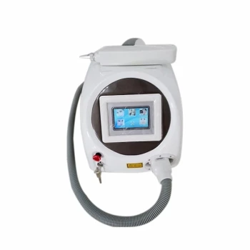
Precision in diagnosis leads to excellence in treatment. BELIS provides premium clinics and high-end salons with professional-grade medical aesthetic equipment, ranging from advanced Pico and Nd:YAG laser systems for scar revision to specialized skin testers that provide the high-definition imaging necessary for scientific evaluation.
Whether you are refining skin texture with Micronedle RF or validating results with our advanced diagnostic tools, BELIS ensures your practice delivers measurable, visible success to your patients.
Ready to upgrade your clinic's diagnostic and treatment capabilities? Contact our experts today to find the perfect system for your practice." Form)"
References
- Noor Taha. The The Combination Effect of Co2 Laser and Topical Growth Factor Solution for Treatment of Atrophic Post-Burn Scar. DOI: 10.32802/asmscj.2022.1215
This article is also based on technical information from Belislaser Knowledge Base .
Related Products
- 22D HIFU Machine Device Facial Machine
- 9D 7D HIFU Vaginal RF Lifting Treatment
- 7D 12D 4D HIFU Machine Device
- IPL SHR+Radio frecuency machine
- 12D HIFU Machine Device for Facial HIFU Treatment
People Also Ask
- What is the mechanism of HIFU devices for facial lifting? Unlock Non-Surgical SMAS Tightening Secrets
- Is swelling a frequent side effect after a HIFU facial? Expert Insights on HIFU Recovery and Safety
- What are the benefits of HIFU machine? Achieve Non-Surgical Skin Lifting & Tightening
- What precautions should be taken when using a High Intensity Focused Ultrasound (HIFU) machine? Expert Safety Guide
- Why is high-precision digital dermatoscopy required for HIFU? Ensure Diagnostic Safety & Precision